Chan Joyce M S, Cheung Maggie S H, Gibbs Richard G J, Bhakoo Kishore K
Department of Surgery, Prince of Wales Hospital, The Chinese University of Hong Kong, Hong Kong SAR, People's Republic of China.
Regional Vascular Unit, St Mary's Hospital, Imperial College Healthcare NHS Trust, Imperial College London, London, UK.
Clin Transl Med. 2017 Dec;6(1):1. doi: 10.1186/s40169-016-0134-1. Epub 2017 Jan 2.
There is currently no clinical imaging technique available to assess the degree of inflammation associated with atherosclerotic plaques. This study aims to develop targeted superparamagnetic particles of iron oxide (SPIO) as a magnetic resonance imaging (MRI) probe for detecting inflamed endothelial cells.
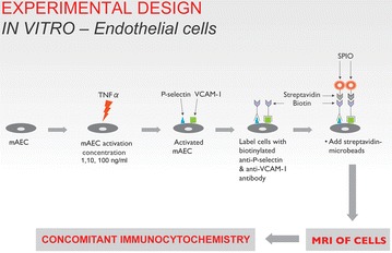
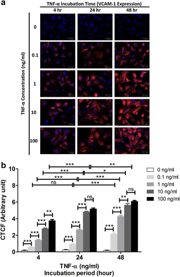
The in vitro study consists of the characterisation and detection of inflammatory markers on activated endothelial cells by immunocytochemistry and MRI using biotinylated anti-P-selectin and anti-VCAM-1 (vascular cell adhesion molecule 1) antibody and streptavidin conjugated SPIO.
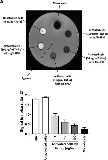
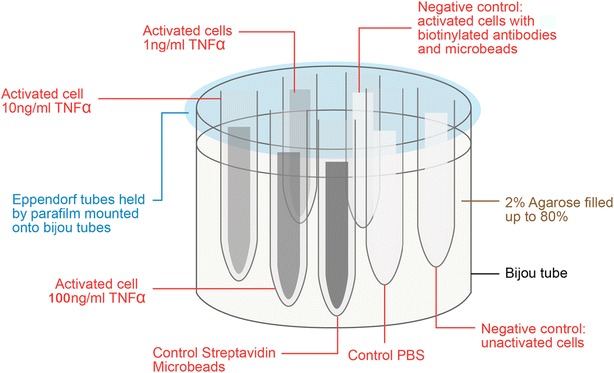
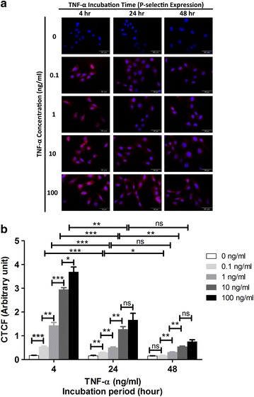
Established an in vitro cellular model of endothelial inflammation induced with TNF-α (tumor necrosis factor alpha). Inflammation of endothelial cells was confirmed with both immunocytochemistry and MRI. These results revealed both a temporal and dose dependent expression of the inflammatory markers, P-selectin and VCAM-1, on exposure to TNF-α.
This study has demonstrated the development of an in vitro model to characterise and detect inflamed endothelial cells by immunocytochemistry and MRI. This will allow the future development of contrast agents and protocols for imaging vascular inflammation in atherosclerosis. This work may form the basis for a translational study to provide clinicians with a novel tool for the in vivo assessment of atherosclerosis.
目前尚无临床成像技术可用于评估与动脉粥样硬化斑块相关的炎症程度。本研究旨在开发靶向超顺磁性氧化铁颗粒(SPIO)作为磁共振成像(MRI)探针,用于检测炎症内皮细胞。
体外研究包括通过免疫细胞化学和MRI,使用生物素化抗P-选择素和抗血管细胞黏附分子1(VCAM-1)抗体以及链霉亲和素偶联的SPIO,对活化内皮细胞上的炎症标志物进行表征和检测。
建立了由肿瘤坏死因子-α(TNF-α)诱导的内皮炎症体外细胞模型。通过免疫细胞化学和MRI证实了内皮细胞炎症。这些结果揭示了炎症标志物P-选择素和VCAM-1在暴露于TNF-α时的时间和剂量依赖性表达。
本研究证明了通过免疫细胞化学和MRI表征和检测炎症内皮细胞的体外模型的开发。这将为未来开发用于动脉粥样硬化血管炎症成像的造影剂和方案提供可能。这项工作可能为转化研究奠定基础,为临床医生提供一种用于体内评估动脉粥样硬化的新型工具。