Cholewka Agnieszka, Szlag Marta, Ślosarek Krzysztof, Białas Brygida
Radiotherapy and Brachytherapy Treatment Planning Department.
Brachytherapy Department, Maria Sklodowska-Curie Memorial Cancer Centre and Institute of Oncology, Gliwice Branch, Gliwice, Poland.
J Contemp Brachytherapy. 2009 Dec;1(4):207-210. Epub 2010 Jan 13.
In this study two different pre-planning methods (2D vs. 3D) were compared in respect to the implant quality as judged by volumetric and dose parameters of the treatment plans. The aim of this work was to evaluate the influence of the imaging modalities used for pre-planning purpose to the treatment plan quality.
Twenty-four patients treated with HDR multicatheter implants were randomly selected for experiment. All patients underwent breast conserving surgery. Flexible catheters were implanted into the breast through the template. Inter-catheter distance, number of planes and catheters were adjusted, in respect to the size and location of the target. Pre-planning was used to evaluate the implant geometry in respect to the target. Needles number and position were modified if necessary. There were two experimental subgroups consisted of 12 patients each. Different pre-planning procedure was employed in each group. In the first group 2D X-ray imaging system was used. In the second one the 3D pre-planning method based on CT was performed. Treatment plans were evaluated with parameters calculated based on dose-volume histograms (DVHs). Volumetric and dose parameters were used for comparison of the dose distribution between the two experimental subgroups.
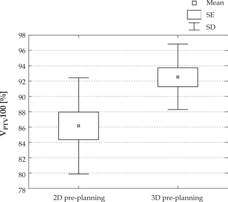
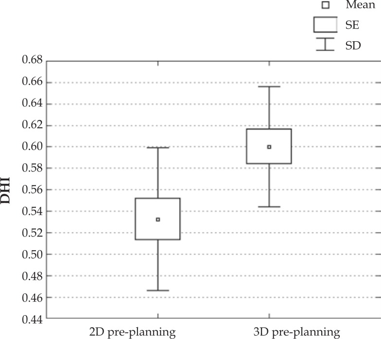
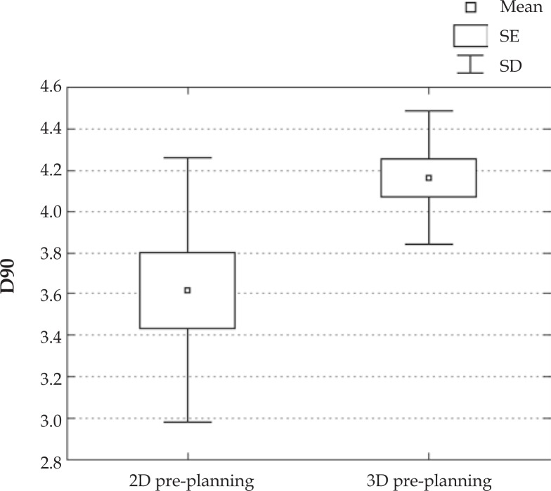
The mean value of target coverage V100 is higher for 3D pre-planning than for 2D (91.7% vs. 86.1%). The dose that covers 90% of the PTV (D90) is also higher for 3D pre-planning than for 2D (4.2 Gy vs. 3.6 Gy). Similar relation can be observed for the values of dose homogeneity index where DHI obtained for 3D pre-planning is 0.60 and 0.53 for 2D. All differences were statistically significant with < 0.05.
Analysis presented in this paper showed that 3D pre-planning method improves the geometrical quality of the implant.
在本研究中,根据治疗计划的体积和剂量参数判断植入质量,对两种不同的预计划方法(二维与三维)进行了比较。这项工作的目的是评估用于预计划目的的成像方式对治疗计划质量的影响。
随机选择24例接受高剂量率多导管植入治疗的患者进行实验。所有患者均接受保乳手术。通过模板将柔性导管植入乳房。根据靶区的大小和位置调整导管间距离、平面数和导管数量。使用预计划来评估植入物相对于靶区的几何形状。必要时修改针的数量和位置。有两个实验亚组,每组12例患者。每组采用不同的预计划程序。第一组使用二维X射线成像系统。第二组采用基于CT的三维预计划方法。使用基于剂量体积直方图(DVH)计算的参数评估治疗计划。使用体积和剂量参数比较两个实验亚组之间的剂量分布。
三维预计划的靶区覆盖率V100的平均值高于二维预计划(91.7%对86.1%)。三维预计划覆盖90%计划靶体积(PTV)的剂量(D90)也高于二维预计划(4.2 Gy对3.6 Gy)。对于剂量均匀性指数值也可观察到类似关系,三维预计划获得的DHI为0.60,二维为0.53。所有差异均具有统计学意义,P<0.05。
本文的分析表明,三维预计划方法提高了植入物的几何质量。