Verfaillie Sander C J, Tijms Betty, Versteeg Adriaan, Benedictus Marije R, Bouwman Femke H, Scheltens Philip, Barkhof Frederik, Vrenken Hugo, van der Flier Wiesje M
Alzheimer Center and Department of Neurology, VU University Medical Center, Amsterdam, The Netherlands.
Department of Radiology and Nuclear Medicine, VU University Medical Center, Amsterdam, The Netherlands.
Alzheimers Dement (Amst). 2016 Nov 19;5:43-52. doi: 10.1016/j.dadm.2016.10.007. eCollection 2016.
We aimed to investigate if thinner cortex of the Alzheimer's disease (AD)-signature region was related to clinical progression in patients with subjective cognitive decline (SCD).
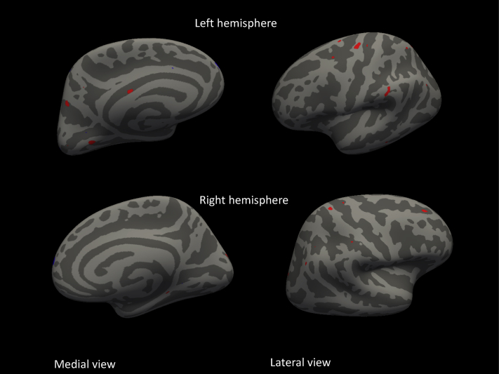
We included 302 SCD patients with clinical follow-up (≥1 year) and three-dimensional T1 magnetic resonance imaging. We estimated AD-signature cortical thickness, consisting of nine frontal, parietal, and temporal gyri and hippocampal volume. We used Cox proportional hazard models (hazard ratios and 95% confidence intervals) to evaluate cortical thickness in relation to clinical progression to mild cognitive impairment (MCI) or dementia.
After a follow-up of the mean (standard deviation) 3 (2) years, 49 patients (16%) showed clinical progression to MCI ( = 32), AD ( = 9), or non-AD dementia ( = 8). Hippocampal volumes, thinner cortex of the AD-signature (hazard ratio [95% confidence interval], 5 [2-17]) and various AD-signature subcomponents were associated with increased risk of clinical progression. Stratified analyses showed that thinner AD-signature cortex was specifically predictive for clinical progression to dementia but not to MCI.
In SCD patients, thinner regional cortex is associated with clinical progression to dementia.
我们旨在研究阿尔茨海默病(AD)特征区域较薄的皮质是否与主观认知下降(SCD)患者的临床进展相关。
我们纳入了302例有临床随访(≥1年)且进行了三维T1磁共振成像的SCD患者。我们估计了AD特征皮质厚度,包括9个额叶、顶叶和颞叶脑回以及海马体积。我们使用Cox比例风险模型(风险比和95%置信区间)来评估皮质厚度与进展为轻度认知障碍(MCI)或痴呆的临床进展之间的关系。
在平均(标准差)3(2)年的随访后,49例患者(16%)出现了进展为MCI(n = 32)、AD(n = 9)或非AD痴呆(n = 8)的临床进展。海马体积、AD特征较薄的皮质(风险比[9%置信区间],5[2 - 17])以及各种AD特征亚成分与临床进展风险增加相关。分层分析表明,AD特征皮质较薄对进展为痴呆具有特异性预测作用,但对进展为MCI则不然。
在SCD患者中,区域皮质较薄与进展为痴呆的临床进展相关。