Francis Willington, Aziz Eid Al Kuwari Maryam A, Ghareep Abdel-Naser, Peyrou Jérôme, Szmigielski Wojciech
Department of Clinical Imaging, Heart Hospital, Hamad Medical Corporation, Doha, Qatar.
Department of Cardiac Imaging, Bordeaux University Hospital, Haut-Lévêque Heart Hospital, Pessac, France.
Pol J Radiol. 2016 Dec 13;81:598-601. doi: 10.12659/PJR.898964. eCollection 2016.
Eosinophilic granulomatosis with polyangiitis (EGPA) is a rare systemic vasculitis with a prevalence rate of seven per million. Cardiac involvement was reported in 20-50%, yet with improved diagnostic methods, the frequency of cardiac involvement is expected to be even higher. It can result in significant morbidity and mortality, accounting for about 50% of death. Cardiac magnetic resonance (CMR) imaging is used to evaluate the myocardium, valves, coronary arteries, pericardium, also to assess cardiac structure and function. Perfusion study allows tissue characterisation with a suggestive pattern of late gadolinium enhancement.
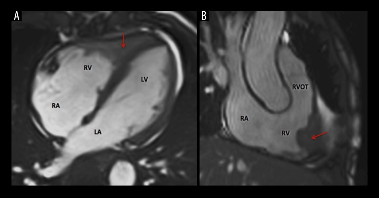
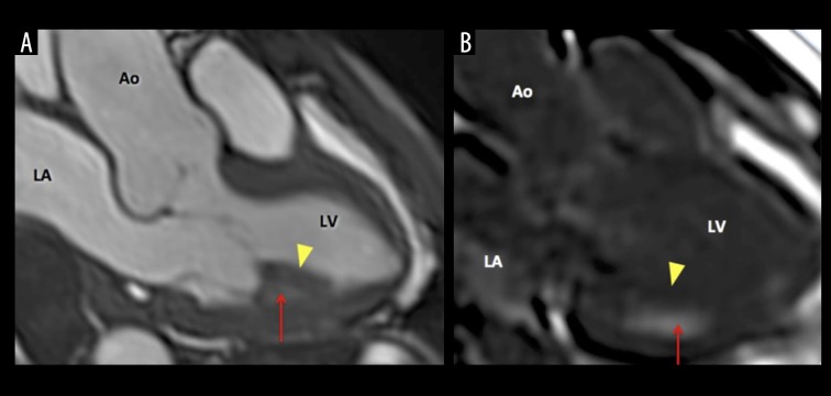
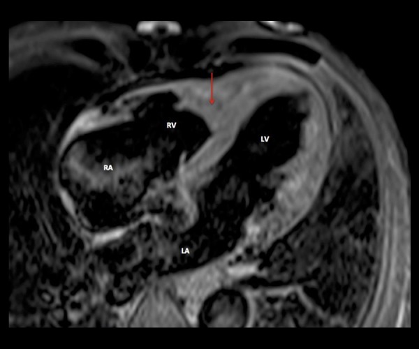
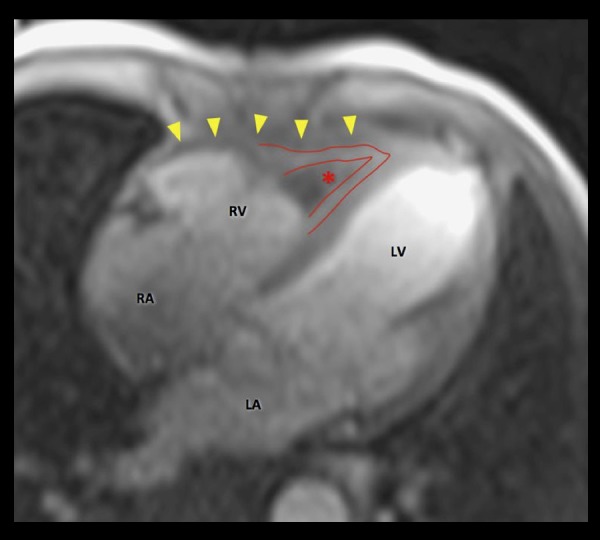
We report a rare case of EGPA in a 54-year-old male patient who presented with fever, sore throat and dizziness. Echocardiography showed a filling defect at the apex of the right ventricle (RV). CMR findings suggested the diagnosis of EGPA by demonstrating an impressive lesion at RV apex with the typical 3-layer appearance and thrombus formation. Post-gadolinium subendocardial hyperenhancement suggested focal involvement at the inferolateral wall of the left ventricle. Computed Tomography (CT) was done to rule out calcific or soft plaques of the coronary arteries, small vessel vasculitis and small aneurysm. CT scan showed a low-attenuation lesion at the inner wall of the right ventricle. In the lungs, bilateral interstitial changes and bilateral cystic bronchiectases were found. Under appropriate treatment, the patient improved clinically.
It is of crucial importance to perform full cardiac imaging that includes CMR even in asymptomatic patients with suspected EGPA, since early identification of cardiac involvement may allow to apply appropriate therapy and full recovery of the patient.
嗜酸性肉芽肿性多血管炎(EGPA)是一种罕见的系统性血管炎,患病率为百万分之七。据报道,心脏受累发生率为20%-50%,然而随着诊断方法的改进,预计心脏受累的频率会更高。它可导致显著的发病率和死亡率,约占死亡人数的50%。心脏磁共振成像(CMR)用于评估心肌、瓣膜、冠状动脉、心包,还可评估心脏结构和功能。灌注研究可通过钆剂延迟强化的特征性表现对组织进行特征性描述。
我们报告了一例54岁男性EGPA罕见病例,该患者表现为发热、咽痛和头晕。超声心动图显示右心室(RV)心尖部有充盈缺损。CMR结果通过显示RV心尖部有典型的三层外观和血栓形成的显著病变提示EGPA诊断。钆剂注射后心内膜下强化提示左心室下外侧壁局灶性受累。进行计算机断层扫描(CT)以排除冠状动脉钙化或软斑块、小血管血管炎和小动脉瘤。CT扫描显示右心室内壁有低密度病变。肺部发现双侧间质性改变和双侧囊状支气管扩张。经适当治疗后,患者临床症状改善。
即使对于疑似EGPA的无症状患者,进行包括CMR在内的全面心脏成像至关重要,因为早期识别心脏受累情况可使患者接受适当治疗并实现完全康复。