Tanthaisong Prapot, Imsoonthornruksa Sumeth, Ngernsoungnern Apichart, Ngernsoungnern Piyada, Ketudat-Cairns Mariena, Parnpai Rangsun
Embryo Technology and Stem Cell Research Center and School of Biotechnology, Suranaree University of Technology, Nakhon Ratchasima, Thailand.
School of Anatomy, Institute of Science, Suranaree University of Technology, Nakhon Ratchasima, Thailand.
PLoS One. 2017 Jan 6;12(1):e0168059. doi: 10.1371/journal.pone.0168059. eCollection 2017.
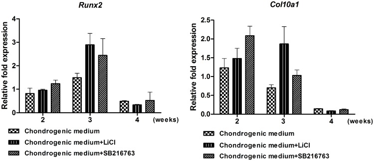
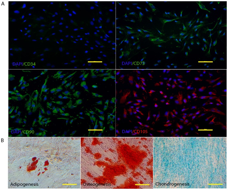
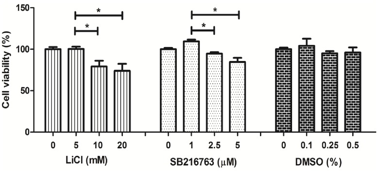
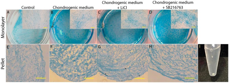
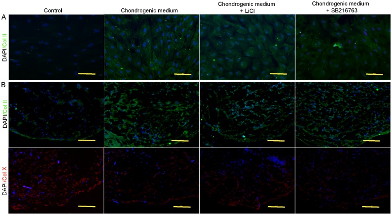
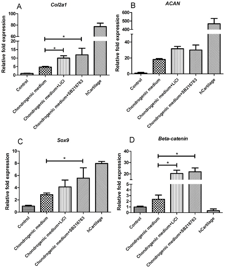
Articular cartilage is an avascular, alymphatic, and aneural system with very low regeneration potential because of its limited capacity for self-repair. Mesenchymal stem cells (MSCs) are the preferred choice for cell-based therapies. Glycogen synthase kinase 3 (GSK-3) inhibitors are compounds that can induce the Wnt signaling pathway, which is involved in chondrogenesis and cartilage development. Here, we investigated the influence of lithium chloride (LiCl) and SB216763 synergistically with TGF-β3 on chondrogenic differentiation in human mesenchymal stem cells derived from Wharton's jelly tissue (hWJ-MSCs). hWJ-MSCs were cultured and chondrogenic differentiation was induced in monolayer and pellet experiments using chondrogenic medium, chondrogenic medium supplemented with LiCl, or SB216763 for 4 weeks. After in vitro differentiation, cultured cells were examined for the expression of Sox9, ACAN, Col2a1, and β-catenin markers. Glycosaminoglycan (GAG) accumulation was also examined by Alcian blue staining. The results indicated that SB216763 was more effective than LiCl as evidenced by a higher up-regulation of the expression of cartilage-specific markers, including Sox9, ACAN, Col2a1 as well as GAG accumulation. Moreover, collagen type II expression was strongly observed in cells cultured in the chondrogenic medium + SB216763 as evidenced by western blot analysis. Both treatments appeared to mediate the Wnt signaling pathway by up-regulating β-catenin gene expression. Further analyses showed that all treatments suppressed the progression of chondrocyte hypertrophy, determined by decreased expression of Col10a1 and Runx2. These results indicate that LiCl and SB216763 are potential candidates for further in vivo therapeutic trials and would be of great importance for cartilage regeneration.
关节软骨是一个无血管、无淋巴管和无神经的系统,由于其自我修复能力有限,再生潜力非常低。间充质干细胞(MSCs)是基于细胞的治疗的首选。糖原合酶激酶3(GSK-3)抑制剂是能够诱导Wnt信号通路的化合物,该信号通路参与软骨形成和软骨发育。在此,我们研究了氯化锂(LiCl)和SB216763与转化生长因子-β3协同作用对源自脐带来源组织的人间充质干细胞(hWJ-MSCs)软骨分化的影响。hWJ-MSCs在单层和微团实验中使用软骨形成培养基、添加LiCl的软骨形成培养基或SB216763培养4周以诱导软骨分化。体外分化后,检测培养细胞中Sox9、ACAN、Col2a1和β-连环蛋白标志物的表达。还通过阿尔辛蓝染色检测糖胺聚糖(GAG)的积累。结果表明,SB216763比LiCl更有效,软骨特异性标志物包括Sox9、ACAN、Col2a1的表达上调以及GAG积累更高证明了这一点。此外,通过蛋白质印迹分析证明,在软骨形成培养基+SB216763中培养的细胞中强烈观察到II型胶原蛋白表达。两种处理似乎都通过上调β-连环蛋白基因表达来介导Wnt信号通路。进一步分析表明,所有处理均通过降低Col10a1和Runx2的表达来抑制软骨细胞肥大的进展。这些结果表明,LiCl和SB216763是进一步进行体内治疗试验的潜在候选物,对软骨再生具有重要意义。