Cortese Francesca, Coppola Gianluca, Di Lenola Davide, Serrao Mariano, Di Lorenzo Cherubino, Parisi Vincenzo, Pierelli Francesco
Department of Medico-Surgical Sciences and Biotechnologies, 'Sapienza' University of Rome Polo Pontino, Corso della Repubblica 79, 04100, Latina, Italy.
G. B. Bietti Foundation IRCCS, Research Unit of Neurophysiology of Vision and Neuro-Ophthalmology, Rome, Italy.
J Headache Pain. 2017 Dec;18(1):2. doi: 10.1186/s10194-016-0712-z. Epub 2017 Jan 6.
Motor-evoked potentials (MEPs) produced by single-pulse transcranial magnetic stimulation (TMS) of the motor cortex can be an objective measure of cortical excitability. Previously, MEP thresholds were found to be normal, increased, or even reduced in patients with migraine. In the present study, we determined whether the level of cortical excitability changes with the time interval from the last migraine attack, thereby accounting for the inconsistencies in previous reports.
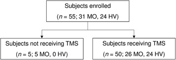
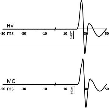
Twenty-six patients with untreated migraine without aura (MO) underwent a MEP study between attacks. Their data were then compared to the MEP data collected from a group of 24 healthy volunteers (HVs). During the experiment, the TMS figure-of-eight coil was positioned over the left motor area. After identifying the resting motor threshold (RMT), we delivered 10 single TMS pulses (rate: 0.1 Hz, intensity: 120% of the RMT) and averaged the resulting MEP amplitudes.
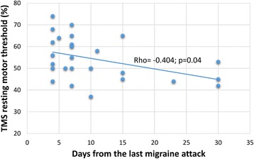
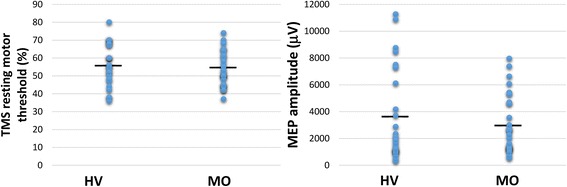
The mean RMTs and MEP amplitudes were not significantly different between the MO and HV groups. In patients with MO, the RMTs were negatively correlated with the number of days elapsed since the last migraine attack (rho = -0.404, p = 0.04).
Our results suggest that the threshold for evoking MEPs is influenced by the proximity of an attack; specifically, the threshold is lower when a long time interval has passed after an attack, and is higher (within the range of normative values) when measured close to an attack. These dynamic RMT variations resemble those we reported previously for visual and somatosensory evoked potentials and may represent time-dependent plastic changes in brain excitability in relation to the migraine cycle.
通过对运动皮层进行单脉冲经颅磁刺激(TMS)产生的运动诱发电位(MEP)可作为皮质兴奋性的客观指标。此前,偏头痛患者的MEP阈值被发现正常、升高甚至降低。在本研究中,我们确定皮质兴奋性水平是否会随着距上次偏头痛发作的时间间隔而变化,从而解释先前报告中的不一致之处。
26例未经治疗的无先兆偏头痛(MO)患者在发作间期接受了MEP研究。然后将他们的数据与从24名健康志愿者(HV)组收集的MEP数据进行比较。在实验过程中,将TMS八字形线圈置于左侧运动区上方。确定静息运动阈值(RMT)后,给予10个单脉冲TMS(频率:0.1Hz,强度:RMT的120%),并对所得MEP波幅进行平均。
MO组和HV组的平均RMT和MEP波幅无显著差异。在MO患者中,RMT与距上次偏头痛发作后的天数呈负相关(rho = -0.404,p = 0.04)。
我们的结果表明,诱发MEP的阈值受发作临近程度的影响;具体而言,发作后经过较长时间间隔时阈值较低,而在接近发作时测量则较高(在正常范围内)。这些动态RMT变化类似于我们先前报道的视觉和体感诱发电位的变化,可能代表与偏头痛周期相关的脑兴奋性随时间的可塑性变化。