Systems Design Engineering, University of Waterloo, Waterloo, N2L3G1, Canada.
Schlegel-University of Waterloo Research Institute for Aging, Waterloo, N2J0E2, Canada.
Sci Rep. 2017 Jan 9;7:40150. doi: 10.1038/srep40150.
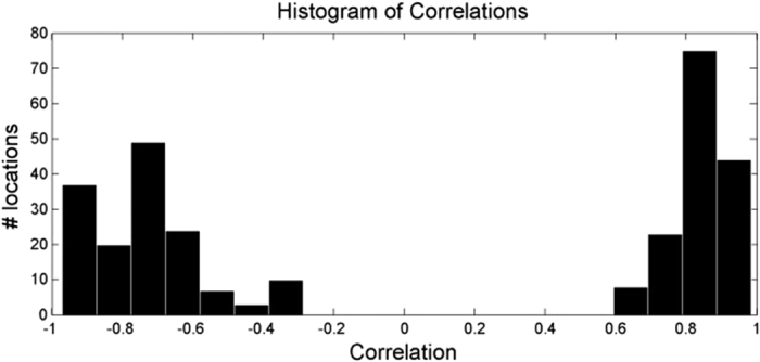
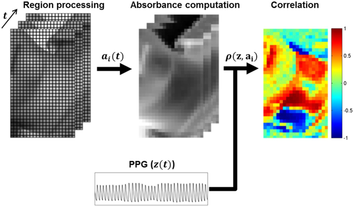
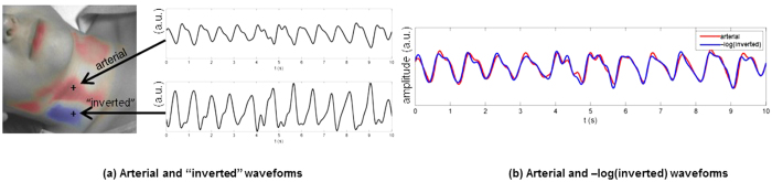
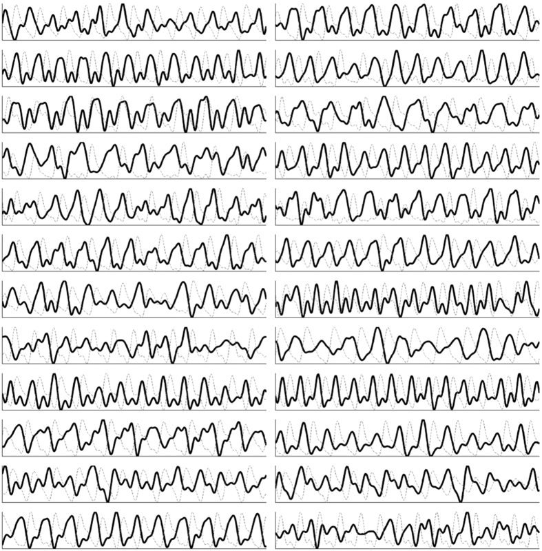
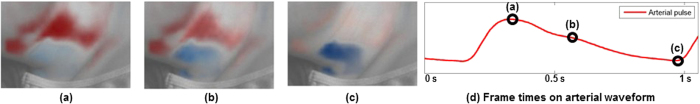
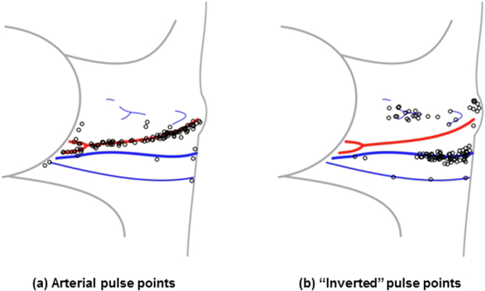
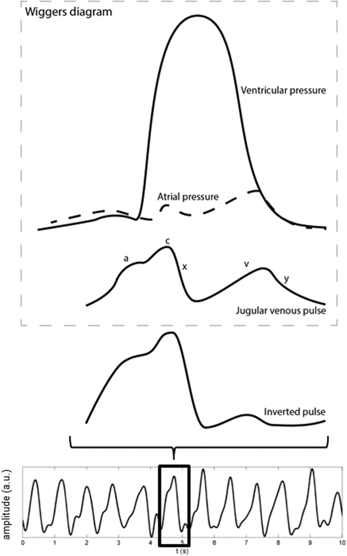
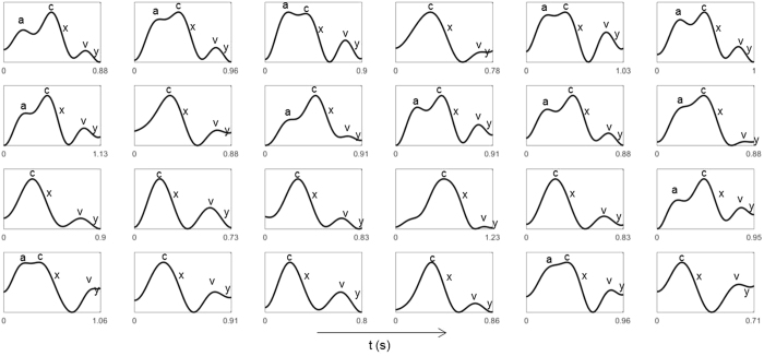
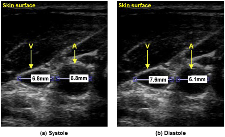
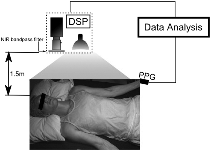
Cardiovascular monitoring is important to prevent diseases from progressing. The jugular venous pulse (JVP) waveform offers important clinical information about cardiac health, but is not routinely examined due to its invasive catheterisation procedure. Here, we demonstrate for the first time that the JVP can be consistently observed in a non-contact manner using a photoplethysmographic imaging system. The observed jugular waveform was strongly negatively correlated to the arterial waveform (r = -0.73 ± 0.17), consistent with ultrasound findings. Pulsatile venous flow was observed over a spatially cohesive region of the neck. Critical inflection points (c, x, v, y waves) of the JVP were observed across all participants. The anatomical locations of the strongest pulsatile venous flow were consistent with major venous pathways identified through ultrasound.
心血管监测对于预防疾病进展很重要。颈静脉搏动(JVP)波形提供了有关心脏健康的重要临床信息,但由于其侵入性的导管插入程序,通常不会进行检查。在这里,我们首次证明,使用光体积描记成像系统可以以非接触的方式一致地观察 JVP。观察到的颈静脉波形与动脉波形呈强烈负相关(r = -0.73 ± 0.17),与超声结果一致。在颈部的一个空间上连贯的区域观察到脉动静脉血流。在所有参与者中都观察到 JVP 的关键拐点(c、x、v、y 波)。脉动静脉血流最强的解剖位置与通过超声确定的主要静脉途径一致。