Qian Xiaoqian, Li Mingyang, Wagner Mary B, Chen Guangping, Song Xiang
Cardiovascular Center, The Fourth Affiliated Hospital, Harbin Medical UniversityHarbin, China; Department of Physiology, Emory University, AtlantaGA, USA.
Cardiovascular Center, The Fourth Affiliated Hospital, Harbin Medical University Harbin, China.
Front Pharmacol. 2016 Dec 20;7:495. doi: 10.3389/fphar.2016.00495. eCollection 2016.
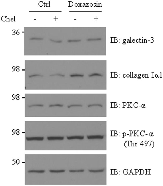
Doxazosin, a drug commonly prescribed for hypertension and prostate disease, increases heart failure risk. However, the underlying mechanism remains unclear. Galectin-3 is an important mediator that plays a pathogenic role in cardiac hypertrophy and heart failure. In the present study, we investigated whether doxazosin could stimulate galectin-3 expression and collagen synthesis in cultured HL-1 cardiomyocytes. We found that doxazosin dose-dependently induced galectin-3 protein expression, with a statistically significant increase in expression with a dose as low as 0.01 μM. Doxazosin upregulated collagen I and α-smooth muscle actin (α-SMA) protein levels and also induced apoptotic protein caspase-3 in HL-1 cardiomyocytes. Although we previously reported that activation of protein kinase C (PKC) stimulates galectin-3 expression, blocking the PKC pathway with the PKC inhibitor chelerythrine did not prevent doxazosin-induced galectin-3 and collagen expression. Consistently, doxazosin treatment did not alter total and phosphorylated PKC. These results suggest that doxazosin-stimulated galectin-3 is independent of PKC pathway. To determine if the α1-adrenergic pathway is involved, we pretreated the cells with the irreversible α-adrenergic receptor blocker phenoxybenzamine and found that doxazosin-stimulated galectin-3 and collagen expression was similar to controls, suggesting that doxazosin acts independently of α1-adrenergic receptor blockade. Collectively, we show a novel effect of doxazosin on cardiomycytes by stimulating heart fibrosis factor galectin-3 expression. The mechanism of action of doxazosin is not mediated through either activation of the PKC pathway or antagonism of α1-adrenergic receptors.
多沙唑嗪是一种常用于治疗高血压和前列腺疾病的药物,它会增加心力衰竭的风险。然而,其潜在机制仍不清楚。半乳糖凝集素-3是一种重要的介质,在心脏肥大和心力衰竭中起致病作用。在本研究中,我们调查了多沙唑嗪是否能刺激培养的HL-1心肌细胞中半乳糖凝集素-3的表达和胶原蛋白合成。我们发现多沙唑嗪剂量依赖性地诱导半乳糖凝集素-3蛋白表达,低至0.01μM的剂量即可使表达有统计学意义的增加。多沙唑嗪上调了HL-1心肌细胞中I型胶原蛋白和α-平滑肌肌动蛋白(α-SMA)的蛋白水平,还诱导了凋亡蛋白半胱天冬酶-3。尽管我们之前报道蛋白激酶C(PKC)的激活会刺激半乳糖凝集素-3的表达,但用PKC抑制剂白屈菜红碱阻断PKC途径并不能阻止多沙唑嗪诱导的半乳糖凝集素-3和胶原蛋白表达。同样,多沙唑嗪处理并未改变总PKC和磷酸化PKC。这些结果表明,多沙唑嗪刺激的半乳糖凝集素-3与PKC途径无关。为了确定α1-肾上腺素能途径是否参与其中,我们用不可逆的α-肾上腺素能受体阻滞剂酚苄明预处理细胞,发现多沙唑嗪刺激的半乳糖凝集素-3和胶原蛋白表达与对照组相似,这表明多沙唑嗪的作用独立于α1-肾上腺素能受体阻断。总的来说,我们通过刺激心脏纤维化因子半乳糖凝集素-3的表达,展示了多沙唑嗪对心肌细胞的一种新作用。多沙唑嗪的作用机制不是通过激活PKC途径或拮抗α1-肾上腺素能受体介导的。