• 文献检索
  • 文档翻译
  • 深度研究
  • 学术资讯
  • Suppr Zotero 插件Zotero 插件
  • 邀请有礼
  • 套餐&价格
  • 历史记录
应用&插件
Suppr Zotero 插件Zotero 插件浏览器插件Mac 客户端Windows 客户端微信小程序
定价
高级版会员购买积分包购买API积分包
服务
文献检索文档翻译深度研究API 文档MCP 服务
关于我们
关于 Suppr公司介绍联系我们用户协议隐私条款
关注我们

Suppr 超能文献

核心技术专利:CN118964589B侵权必究
粤ICP备2023148730 号-1Suppr @ 2026

文献检索

告别复杂PubMed语法,用中文像聊天一样搜索,搜遍4000万医学文献。AI智能推荐,让科研检索更轻松。

立即免费搜索

文件翻译

保留排版,准确专业,支持PDF/Word/PPT等文件格式,支持 12+语言互译。

免费翻译文档

深度研究

AI帮你快速写综述,25分钟生成高质量综述,智能提取关键信息,辅助科研写作。

立即免费体验

健康人体脊髓节段直径综述。

A Review of the Segmental Diameter of the Healthy Human Spinal Cord.

作者信息

Frostell Arvid, Hakim Ramil, Thelin Eric Peter, Mattsson Per, Svensson Mikael

机构信息

Department of Clinical Neuroscience, Karolinska Institutet , Stockholm , Sweden.

Department of Clinical Neuroscience, Karolinska Institutet, Stockholm, Sweden; Department of Neurosurgery, Karolinska University Hospital, Stockholm, Sweden.

出版信息

Front Neurol. 2016 Dec 23;7:238. doi: 10.3389/fneur.2016.00238. eCollection 2016.

DOI:10.3389/fneur.2016.00238
PMID:28066322
原文链接:https://pmc.ncbi.nlm.nih.gov/articles/PMC5179522/
Abstract

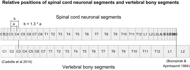
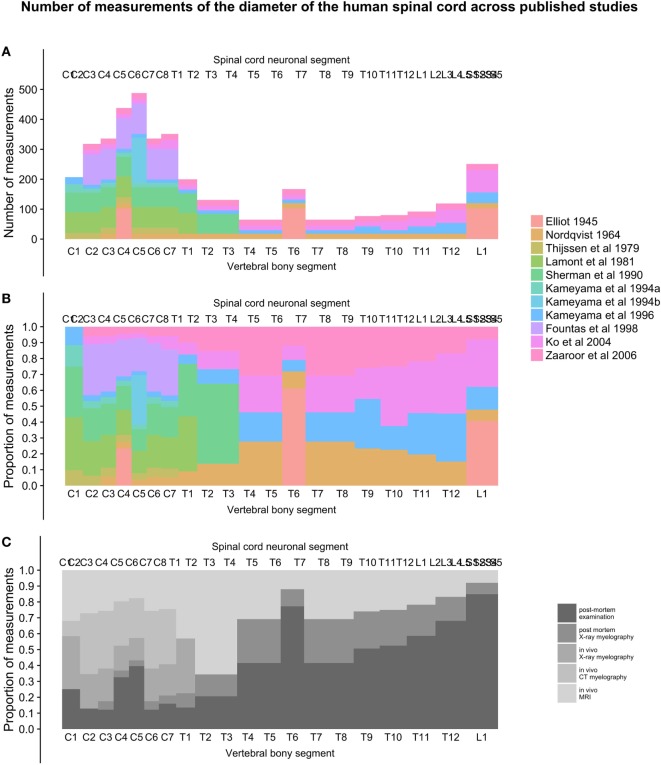
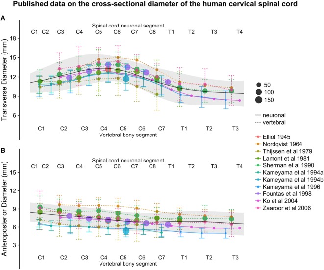
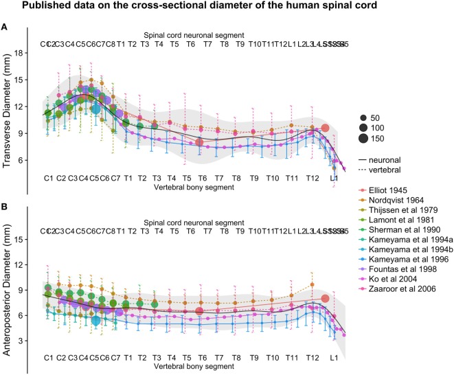
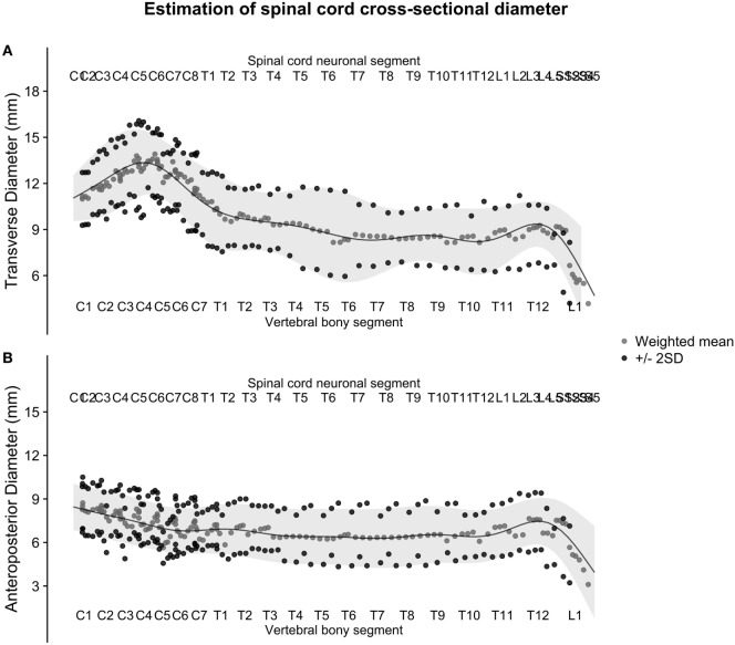
Knowledge of the average size and variability of the human spinal cord can be of importance when treating pathological conditions in the spinal cord. Data on healthy human spinal cord morphometrics have been published for more than a century using different techniques of measurements, but unfortunately, comparison of results from different studies is difficult because of the different anatomical landmarks used as reference points along the craniocaudal axis for the measurements. The aim of this review was to compute population estimates of the transverse and anteroposterior diameter of the human spinal cord by comparing and combining previously published data on a normalized craniocaudal axis. We included 11 studies presenting measurements of spinal cord cross-sectional diameters, with a combined sample size ranging from 15 to 488 subjects, depending on spinal cord level. Based on five published studies presenting data on the lengths of the segments of the spinal cord and vertebral column, we calculated the relative positions of all spinal cord neuronal segments and vertebral bony segments and mapped measurements of spinal cord size to a normalized craniocaudal axis. This mapping resulted in better alignment between studies and allowed the calculation of weighted averages and standard deviations (SDs) along the spinal cord. These weighted averages were smoothed using a generalized additive model to yield continuous population estimates for transverse and anteroposterior diameter and associated SDs. The spinal cord had the largest transverse diameter at spinal cord neuronal segment C5 (13.3 ± 2.2), decreased to segment T8 (8.3 ± 2.1), and increased slightly again to 9.4 ± 1.5 at L3. The anteroposterior diameter showed less variation in size along the spinal cord at C5 (7.4 ± 1.6), T8 (6.3 ± 2.0), and L3 (7.5 ± 1.6). All estimates are presented in millimeters ± 2 SDs. We conclude that segmental transverse and anteroposterior diameters of the healthy human spinal cord from different published sources can be combined on a normalized craniocaudal axis and yield meaningful population estimates. These estimates could be useful in routine management of patients with neurodegenerative diseases as well as for clinical research and experimental applications aimed at surgical spinal cord repair.

摘要

在治疗脊髓疾病时,了解人类脊髓的平均大小和变异性可能具有重要意义。关于健康人类脊髓形态计量学的数据已经使用不同的测量技术发表了一个多世纪,但不幸的是,由于在测量中沿头尾轴用作参考点的解剖标志不同,不同研究结果的比较很困难。本综述的目的是通过比较和合并先前发表的关于标准化头尾轴的数据,计算人类脊髓横径和前后径的总体估计值。我们纳入了11项呈现脊髓横截面积测量值的研究,根据脊髓节段水平,合并样本量从15至488名受试者不等。基于5项发表的关于脊髓节段和脊柱长度数据的研究,我们计算了所有脊髓神经节段和椎骨节段的相对位置,并将脊髓大小的测量值映射到标准化的头尾轴上。这种映射使得各研究之间的对齐更好,并允许计算沿脊髓的加权平均值和标准差(SD)。使用广义相加模型对这些加权平均值进行平滑处理,以得出横径和前后径的连续总体估计值以及相关的SD。脊髓在脊髓神经节段C5处横径最大(13.3±2.2),到节段T8时减小至(8.3±2.1),在L3处又略有增加至9.4±1.5。前后径在脊髓C5(7.4±1.6)、T8(6.3±2.0)和L3(7.5±1.6)处沿脊髓的大小变化较小。所有估计值均以毫米±2SD表示。我们得出结论,来自不同已发表来源的健康人类脊髓节段横径和前后径可以在标准化的头尾轴上合并,并得出有意义的总体估计值。这些估计值对于神经退行性疾病患者的常规管理以及旨在脊髓手术修复的临床研究和实验应用可能有用。

https://cdn.ncbi.nlm.nih.gov/pmc/blobs/1d64/5179522/4b115a55b126/fneur-07-00238-g006.jpg
https://cdn.ncbi.nlm.nih.gov/pmc/blobs/1d64/5179522/bc647e4a74ec/fneur-07-00238-g001.jpg
https://cdn.ncbi.nlm.nih.gov/pmc/blobs/1d64/5179522/4fa9770ca480/fneur-07-00238-g002.jpg
https://cdn.ncbi.nlm.nih.gov/pmc/blobs/1d64/5179522/58dffc33eafd/fneur-07-00238-g003.jpg
https://cdn.ncbi.nlm.nih.gov/pmc/blobs/1d64/5179522/d7890f1d18a0/fneur-07-00238-g004.jpg
https://cdn.ncbi.nlm.nih.gov/pmc/blobs/1d64/5179522/4aa85035965e/fneur-07-00238-g005.jpg
https://cdn.ncbi.nlm.nih.gov/pmc/blobs/1d64/5179522/4b115a55b126/fneur-07-00238-g006.jpg

相似文献

1
A Review of the Segmental Diameter of the Healthy Human Spinal Cord.健康人体脊髓节段直径综述。
Front Neurol. 2016 Dec 23;7:238. doi: 10.3389/fneur.2016.00238. eCollection 2016.
2
Morphometrics of the entire human spinal cord and spinal canal measured from in vivo high-resolution anatomical magnetic resonance imaging.通过活体高分辨率解剖磁共振成像测量整个人类脊髓和椎管的形态学指标。
Spine (Phila Pa 1976). 2014 Feb 15;39(4):E262-9. doi: 10.1097/BRS.0000000000000125.
3
Relatively Large Cervical Spinal Cord for Spinal Canal is a Risk factor for Development of Cervical Spinal Cord Compression: A Cross-Sectional Study of 1211 Subjects.脊髓管径相对较大是发生颈脊髓压迫症的一个危险因素:一项对1211名受试者的横断面研究
Spine (Phila Pa 1976). 2016 Mar;41(6):E342-8. doi: 10.1097/BRS.0000000000001255.
4
Cervical spinal cord--smaller than considered?颈脊髓——比预想的小?
Spine (Phila Pa 1976). 1998 Jul 15;23(14):1513-6. doi: 10.1097/00007632-199807150-00001.
5
Morphometry of the normal cadaveric cervical spinal cord.正常尸体颈脊髓的形态测量学
Spine (Phila Pa 1976). 1994 Sep 15;19(18):2077-81. doi: 10.1097/00007632-199409150-00013.
6
Distribution of sympathetic preganglionic neurons innervating the kidney in the rat: PRV transneuronal tracing and serial reconstruction.大鼠中支配肾脏的交感神经节前神经元的分布:PRV跨神经元追踪及连续重建
Auton Neurosci. 2002 Jan 10;95(1-2):57-70. doi: 10.1016/s1566-0702(01)00356-3.
7
[Morphometry of the normal cadaveric cervical spinal cord].[正常尸体颈段脊髓的形态测量学]
Rinsho Shinkeigaku. 1992 Nov;32(11):1203-7.
8
Congenital lumbar spinal stenosis: a prospective, control-matched, cohort radiographic analysis.先天性腰椎管狭窄症:一项前瞻性、对照匹配队列影像学分析。
Spine J. 2005 Nov-Dec;5(6):615-22. doi: 10.1016/j.spinee.2005.05.385.
9
Morphological and functional electrophysiological evidence of relative spinal cord tethering in adolescent idiopathic scoliosis.青少年特发性脊柱侧凸中相对脊髓拴系的形态学和功能性电生理证据。
Spine (Phila Pa 1976). 2008 Mar 15;33(6):673-80. doi: 10.1097/BRS.0b013e318166aa58.
10
Diametral measurements of the upper spinal cord for stereotactic pain procedures: experimental and clinical study.用于立体定向疼痛治疗的上脊髓直径测量:实验与临床研究
Surg Neurol. 1995 May;43(5):478-82; discussion 482-3. doi: 10.1016/0090-3019(95)80093-v.

引用本文的文献

1
Rootlets-based registration to the PAM50 spinal cord template.基于神经根丝的PAM50脊髓模板配准
Imaging Neurosci (Camb). 2025 Aug 26;3. doi: 10.1162/IMAG.a.123. eCollection 2025.
2
Fragile X-associated tremor/ataxia syndrome: a case report with atypical spinal cord atrophy and altered somatosensory evoked potentials.脆性X相关震颤/共济失调综合征:一例伴有非典型脊髓萎缩和体感诱发电位改变的病例报告
J Neurol. 2025 Aug 28;272(9):595. doi: 10.1007/s00415-025-13333-4.
3
Towards personalized mapping through lumbosacral spinal cord task fMRI.

本文引用的文献

1
Concise Review: Bridging the Gap: Novel Neuroregenerative and Neuroprotective Strategies in Spinal Cord Injury.简要综述:弥合差距:脊髓损伤中的新型神经再生和神经保护策略
Stem Cells Transl Med. 2016 Jul;5(7):914-24. doi: 10.5966/sctm.2015-0381. Epub 2016 Apr 29.
2
Spinal cord MRI in multiple sclerosis--diagnostic, prognostic and clinical value.多发性硬化症的脊髓 MRI——诊断、预后和临床价值。
Nat Rev Neurol. 2015 Jun;11(6):327-38. doi: 10.1038/nrneurol.2015.80. Epub 2015 May 26.
3
Characterizing the location of spinal and vertebral levels in the human cervical spinal cord.
通过腰骶部脊髓任务功能磁共振成像实现个性化映射
Imaging Neurosci (Camb). 2025 Jan 23;3. doi: 10.1162/imag_a_00455. eCollection 2025.
4
Reliability of task-based fMRI in the dorsal horn of the human spinal cord.基于任务的功能磁共振成像在人类脊髓背角的可靠性
Imaging Neurosci (Camb). 2024 Aug 22;2. doi: 10.1162/imag_a_00273. eCollection 2024.
5
Lumbosacral spinal cord functional connectivity at rest: From feasibility to reliability.静息状态下腰骶脊髓的功能连接:从可行性到可靠性
Imaging Neurosci (Camb). 2024 Sep 5;2. doi: 10.1162/imag_a_00286. eCollection 2024.
6
parcellation of the human spinal cord functional architecture.人类脊髓功能结构的分区
Imaging Neurosci (Camb). 2024 Jan 11;2. doi: 10.1162/imag_a_00059. eCollection 2024.
7
A database of the healthy human spinal cord morphometry in the PAM50 template space.PAM50模板空间中健康人类脊髓形态测量的数据库。
Imaging Neurosci (Camb). 2024 Feb 2;2. doi: 10.1162/imag_a_00075. eCollection 2024.
8
Functional magnetic resonance imaging of the lumbosacral cord during a lower extremity motor task.下肢运动任务期间腰骶脊髓的功能磁共振成像
Imaging Neurosci (Camb). 2024 Jul 15;2. doi: 10.1162/imag_a_00227. eCollection 2024.
9
Cerebro-spinal somatotopic organization uncovered through functional connectivity mapping.通过功能连接图谱揭示的脑脊髓躯体定位组织
Imaging Neurosci (Camb). 2024 Sep 5;2. doi: 10.1162/imag_a_00284. eCollection 2024.
10
Automatic segmentation of the spinal cord nerve rootlets.脊髓神经根丝的自动分割
Imaging Neurosci (Camb). 2024 Jul 2;2. doi: 10.1162/imag_a_00218. eCollection 2024.
确定人类颈脊髓中脊髓节段和椎体水平的位置。
AJNR Am J Neuroradiol. 2015 Apr;36(4):803-10. doi: 10.3174/ajnr.A4192. Epub 2014 Dec 18.
4
Method for simultaneous voxel-based morphometry of the brain and cervical spinal cord area measurements using 3D-MDEFT.使用 3D-MDEFT 进行基于体素的脑和颈椎脊髓区域测量的同步形态测量方法。
J Magn Reson Imaging. 2010 Nov;32(5):1242-7. doi: 10.1002/jmri.22340.
5
Cross-sectional diameters and areas of the human spinal cord.人类脊髓的横截面积和直径
Anat Rec. 1945 Nov;93:287-93. doi: 10.1002/ar.1090930306.
6
Morphological study of the spinal canal content for subarachnoid endoscopy.用于蛛网膜下腔内镜检查的椎管内容物的形态学研究
Minim Invasive Neurosurg. 2006 Aug;49(4):220-6. doi: 10.1055/s-2006-948000.
7
Gross quantitative measurements of spinal cord segments in human.人体脊髓节段的大体定量测量。
Spinal Cord. 2004 Jan;42(1):35-40. doi: 10.1038/sj.sc.3101538.
8
THE SAGITTAL DIAMETER OF THE SPINAL CORD AND SUBARACHNOID SPACE IN DIFFERENT AGE GROUPS. A ROENTGENOGRAPHIC POST-MORTEM STUDY.不同年龄组脊髓和蛛网膜下腔的矢状径:一项X线尸检研究
Acta Radiol Diagn (Stockh). 1964:SUPPL 227:1-96.
9
Dimensions of the cervical vertebrae.颈椎的尺寸。
Anat Rec. 1955 Aug;122(4):603-9. doi: 10.1002/ar.1091220409.
10
Spinal-cord injury.脊髓损伤
Lancet. 2002 Feb 2;359(9304):417-25. doi: 10.1016/S0140-6736(02)07603-1.