Shi Yanyan, Wei Ying, Zhang Ting, Zhang Jing, Wang Ye, Ding Shigang
Research Center of Clinical Epidemiology, Peking University Third Hospital, Beijing 100191, China.
Department of Gastroenterology, Peking University Third Hospital, Beijing 100191, China.
Gastroenterol Res Pract. 2016;2016:9638963. doi: 10.1155/2016/9638963. Epub 2016 Dec 14.
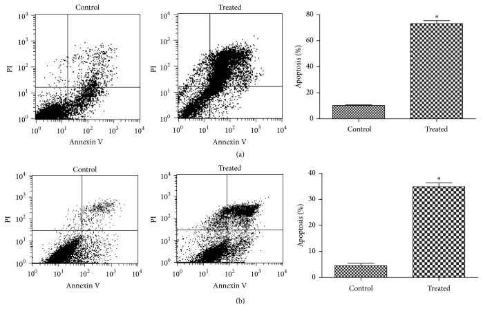
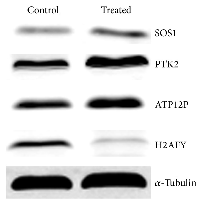
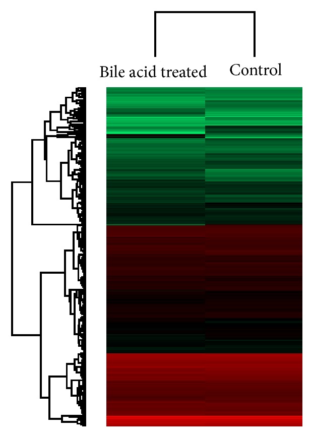
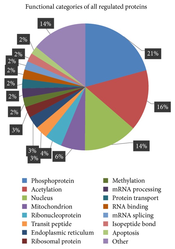
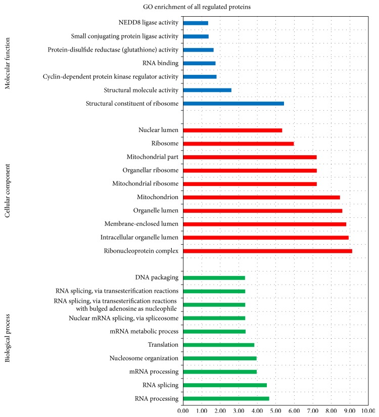
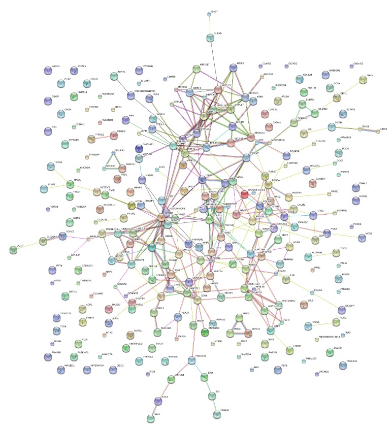
Pathologic duodenogastric reflux can induce or aggravate gastritis because of the presence of bile acids. Bile reflux has been generally considered to be associated with intestinal metaplasia and gastric cancer. However, the pathogenic mechanisms of the effects of bile acids on gastric mucosa are still unknown. To explore the mechanisms by which bile acids induce gastric mucosal lesions, we examined cell apoptosis in the gastric epithelial cell line GES-1 and investigated the changes in protein profiles of GES-1 cells in response to a bile acid deoxycholic acid using a proteomics approach. Changes in the profiles of the differently expressed proteins were analyzed using the DAVID and STRING programs. We found apoptosis was significantly induced in GES-1 cells by deoxycholic acid. Using liquid chromatographic/tandem mass spectrometric (LC-MS/MS) methods, 134 upregulated proteins and 214 downregulated proteins were identified in the bile acid treated GES-1 cells. Bioinformatics analysis revealed the interactions and signaling networks of these differentially expressed proteins. These findings may improve the understanding of the molecular mechanisms underlying the pathogenicity of bile acids on gastric mucosa.
病理性十二指肠-胃反流可因胆汁酸的存在而诱发或加重胃炎。胆汁反流通常被认为与肠化生和胃癌有关。然而,胆汁酸对胃黏膜作用的致病机制仍不清楚。为了探究胆汁酸诱导胃黏膜损伤的机制,我们检测了胃上皮细胞系GES-1中的细胞凋亡,并使用蛋白质组学方法研究了GES-1细胞对胆汁酸脱氧胆酸反应时蛋白质谱的变化。使用DAVID和STRING程序分析差异表达蛋白质谱的变化。我们发现脱氧胆酸可显著诱导GES-1细胞凋亡。采用液相色谱/串联质谱(LC-MS/MS)方法,在经胆汁酸处理的GES-1细胞中鉴定出134种上调蛋白和214种下调蛋白。生物信息学分析揭示了这些差异表达蛋白质的相互作用和信号网络。这些发现可能有助于增进对胆汁酸对胃黏膜致病性的分子机制的理解。