Junka Adam, Szymczyk Patrycja, Ziółkowski Grzegorz, Karuga-Kuzniewska Ewa, Smutnicka Danuta, Bil-Lula Iwona, Bartoszewicz Marzenna, Mahabady Susan, Sedghizadeh Parish Paymon
Department of Pharmaceutical Microbiology and Parasitology, Wroclaw Medical University, Wroclaw, Poland.
Center for Advanced Manufacturing Technologies (CAMT/FPC), Faculty of Mechanical Engineering, Wroclaw University of Technology, Wroclaw, Poland.
PLoS One. 2017 Jan 11;12(1):e0169565. doi: 10.1371/journal.pone.0169565. eCollection 2017.
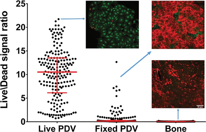
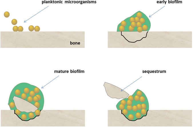
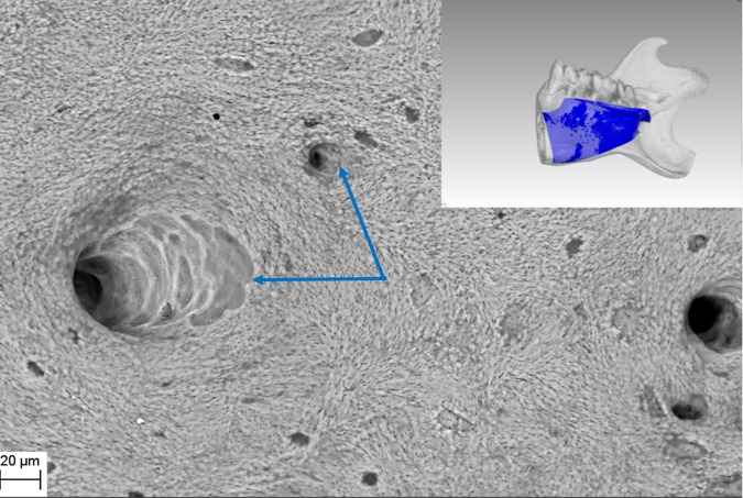
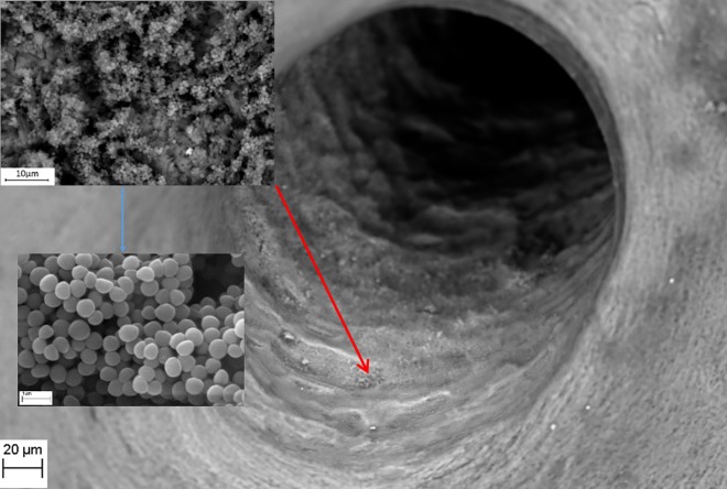
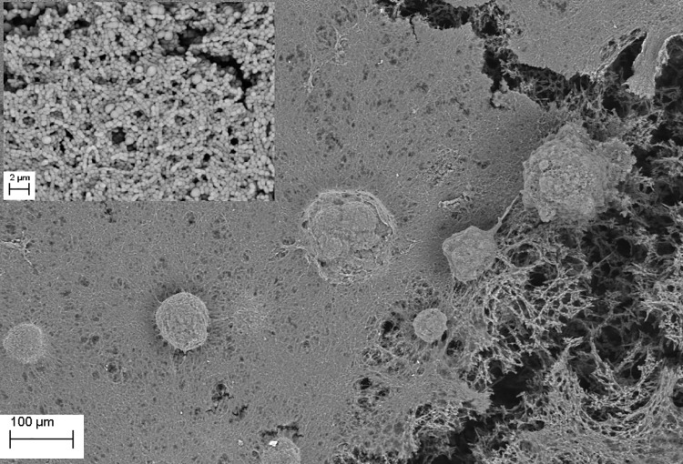
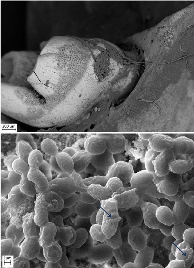
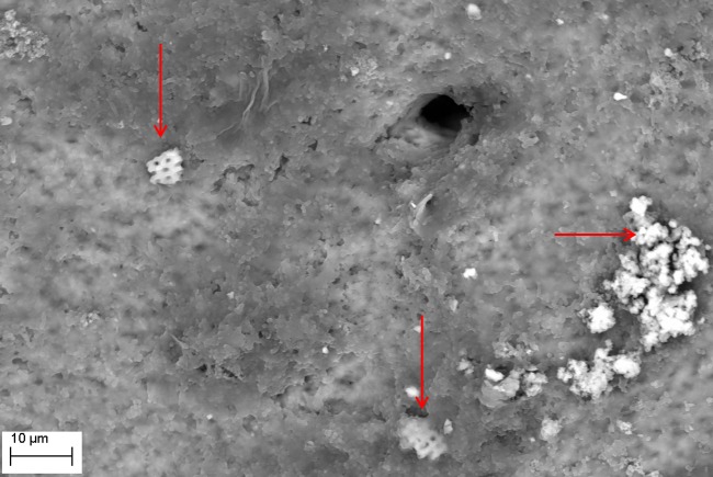
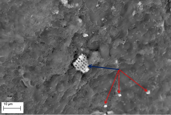
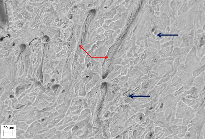
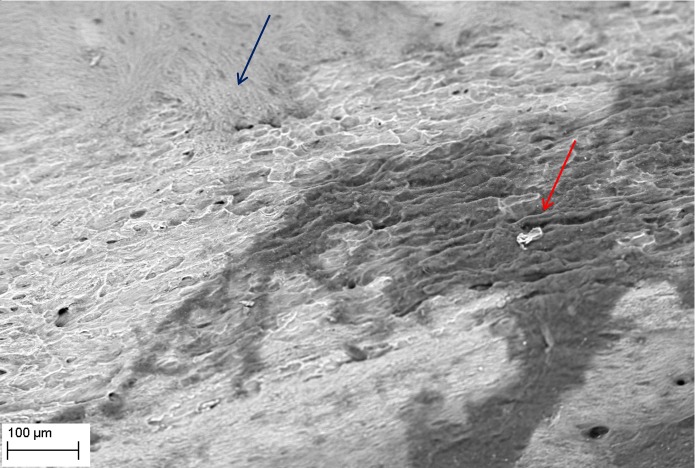
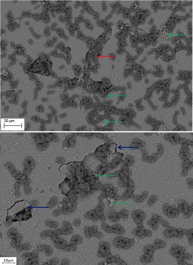
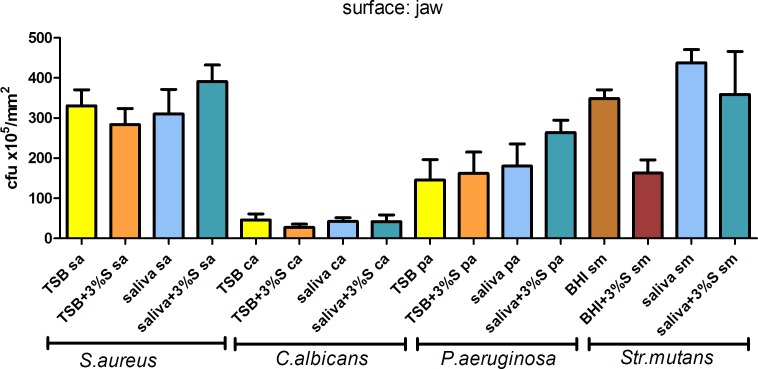
Bone infections are a significant public health burden associated with morbidity and mortality in patients. Microbial biofilm pathogens are the causative agents in chronic osteomyelitis. Research on the pathogenesis of osteomyelitis has focused on indirect bone destruction by host immune cells and cytokines secondary to microbial insult. Direct bone resorption by biofilm pathogens has not yet been seriously considered. In this study, common osteomyelitis pathogens (Staphylococcus aureus, Pseudomonas aeruginosa, Candida albicans, and Streptococcus mutans) were grown as biofilms in multiple in vitro and ex vivo experiments to analyze quantitative and qualitative aspects of bone destruction during infection. Pathogens were grown as single or mixed species biofilms on the following substrates: hydroxyapatite, rat jawbone, or polystyrene wells, and in various media. Biofilm growth was evaluated by scanning electron microscopy and pH levels were monitored over time. Histomorphologic and quantitative effects of biofilms on tested substrates were analyzed by microcomputed tomography and quantitative cultures. All tested biofilms demonstrated significant damage to bone. Scanning electron microscopy indicated that all strains formed mature biofilms within 7 days on all substrate surfaces regardless of media. Experimental conditions impacted pH levels, although this had no impact on biofilm growth or bone destruction. Presence of biofilm led to bone dissolution with a decrease of total volume by 20.17±2.93% upon microcomputed tomography analysis, which was statistically significant as compared to controls (p <0.05, ANOVA). Quantitative cultures indicated that media and substrate did not impact biofilm formation (Kruskall-Wallis test, post-hoc Dunne's test; p <0.05). Overall, these results indicate that biofilms associated with osteomyelitis have the ability to directly resorb bone. These findings should lead to a more complete understanding of the etiopathogenesis of osteomyelitis, where direct bone resorption by biofilm is considered in addition to the well-known osteoclastic and host cell destruction of bone.
骨感染是一种与患者发病率和死亡率相关的重大公共卫生负担。微生物生物膜病原体是慢性骨髓炎的致病因素。骨髓炎发病机制的研究主要集中在宿主免疫细胞和微生物损伤继发的细胞因子对骨的间接破坏上。生物膜病原体对骨的直接吸收尚未得到认真考虑。在本研究中,常见的骨髓炎病原体(金黄色葡萄球菌、铜绿假单胞菌、白色念珠菌和变形链球菌)在多个体外和体内实验中形成生物膜,以分析感染期间骨破坏的定量和定性方面。病原体在以下底物上形成单一或混合物种生物膜:羟基磷灰石、大鼠颌骨或聚苯乙烯孔,并在各种培养基中培养。通过扫描电子显微镜评估生物膜生长,并随时间监测pH值水平。通过微计算机断层扫描和定量培养分析生物膜对测试底物的组织形态学和定量影响。所有测试的生物膜均对骨造成了显著损伤。扫描电子显微镜表明,所有菌株在7天内在所有底物表面形成了成熟的生物膜,无论培养基如何。实验条件影响pH值水平,尽管这对生物膜生长或骨破坏没有影响。生物膜的存在导致骨溶解,微计算机断层扫描分析显示总体积减少20.17±2.93%,与对照组相比具有统计学意义(p<0.05,方差分析)。定量培养表明,培养基和底物不影响生物膜形成(Kruskal-Wallis检验,事后Dunne检验;p<0.05)。总体而言,这些结果表明,与骨髓炎相关的生物膜具有直接吸收骨的能力。这些发现应该会使人们对骨髓炎的病因发病机制有更全面的了解,其中除了众所周知的破骨细胞和宿主细胞对骨的破坏外,还应考虑生物膜对骨的直接吸收。