Nishimura Chiaki
Faculty of Pharmaceutical Sciences, Teikyo Heisei University.
Proc Jpn Acad Ser B Phys Biol Sci. 2017;93(1):10-27. doi: 10.2183/pjab.93.002.
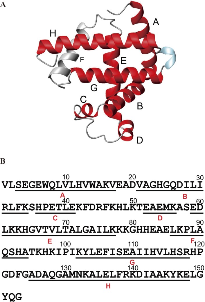
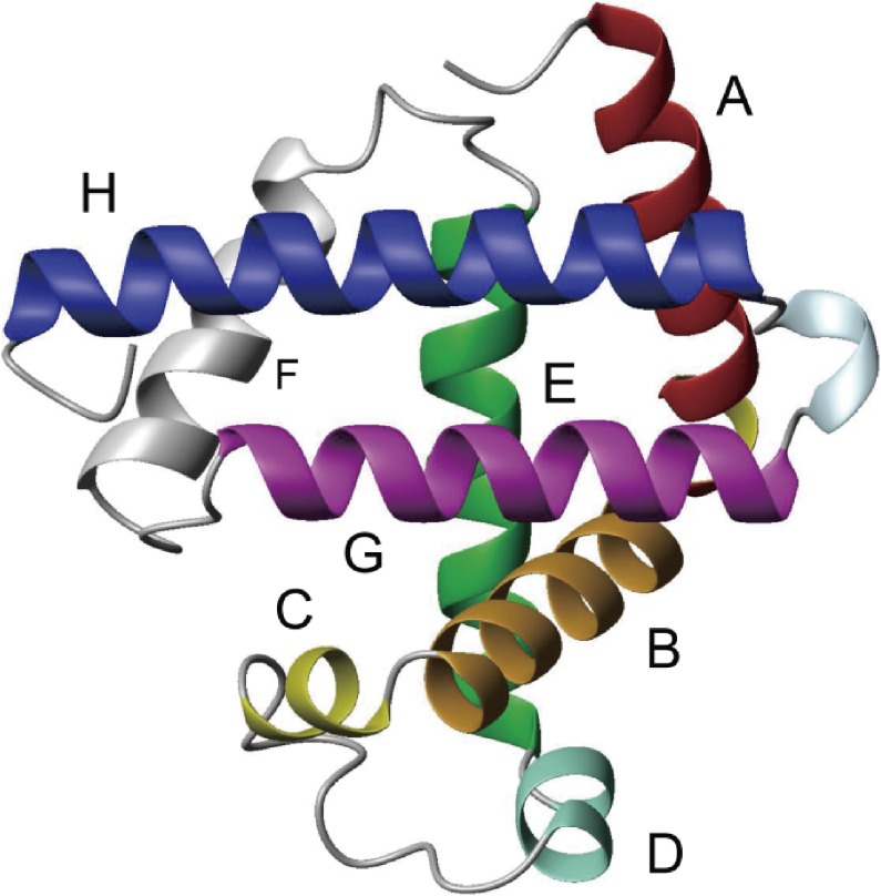
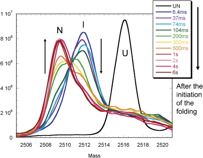
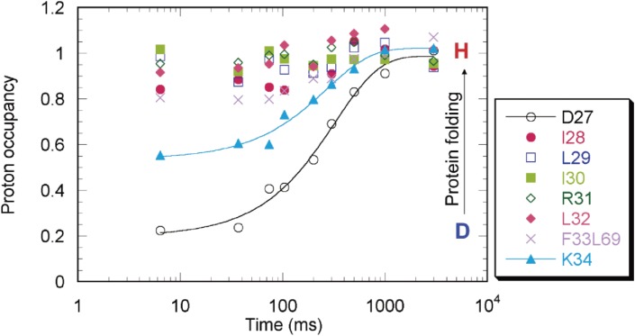
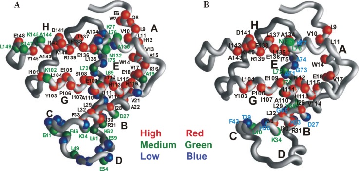
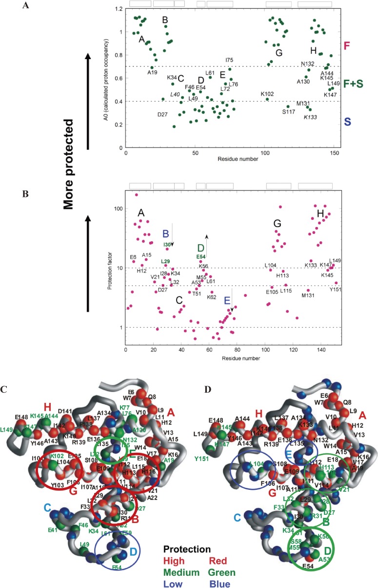
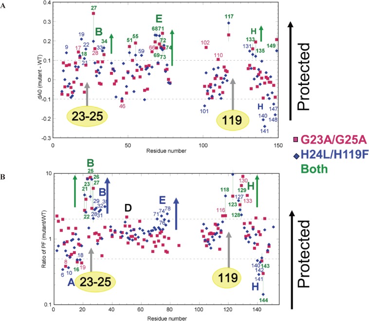
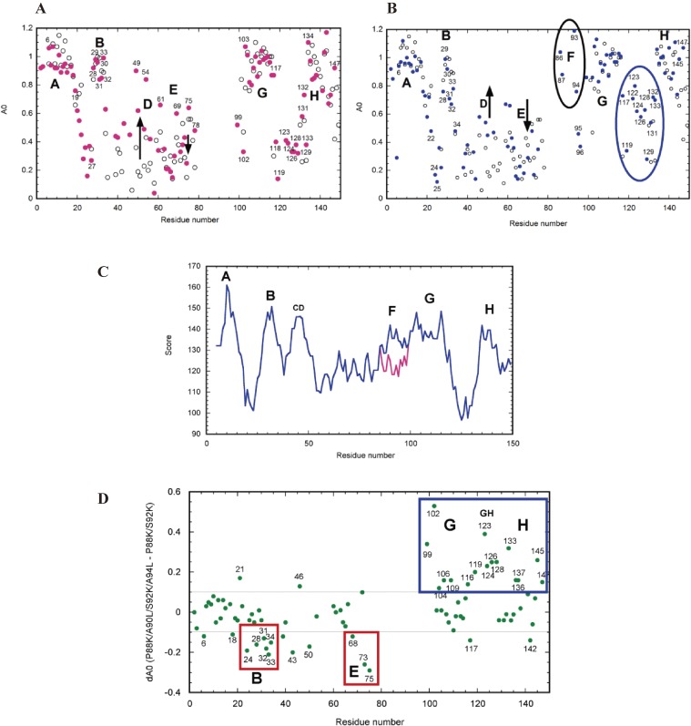
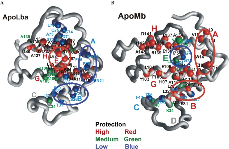
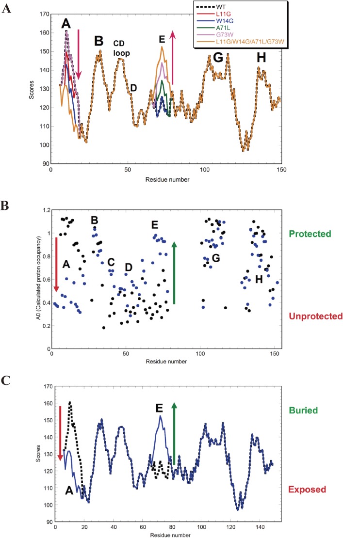
The structures of apomyoglobin folding intermediates have been widely analyzed using physical chemistry methods including fluorescence, circular dichroism, small angle X-ray scattering, NMR, mass spectrometry, and rapid mixing. So far, at least two intermediates (on sub-millisecond- and millisecond-scales) have been demonstrated for apomyoglobin folding. The combination of pH-pulse labeling and NMR is a useful tool for analyzing the kinetic intermediates at the atomic level. Its use has revealed that the latter-phase kinetic intermediate of apomyoglobin (6 ms) was composed of helices A, B, G and H, whereas the equilibrium intermediate, called the pH 4 molten-globule intermediate, was composed mainly of helices A, G and H. The improved strategy for the analysis of the kinetic intermediate was developed to include (1) the dimethyl sulfoxide method, (2) data processing with the various labeling times, and (3) a new in-house mixer. Particularly, the rapid mixing revealed that helices A and G were significantly more protected at the earlier stage (400 µs) of the intermediate (former-phase intermediate) than the other helices. Mutation studies, where each hydrophobic residue was replaced with an alanine in helices A, B, E, F, G and H, indicated that both non-native and native-like structures exist in the latter-phase folding intermediate. The N-terminal part of helix B is a weak point in the intermediate, and the docking of helix E residues to the core of the A, B, G and H helices was interrupted by a premature helix B, resulting in the accumulation of the intermediate composed of helices A, B, G and H. The prediction-based protein engineering produced important mutants: Helix F in a P88K/A90L/S92K/A94L mutant folded in the latter-phase intermediate, although helix F in the wild type does not fold even at the native state. Furthermore, in the L11G/W14G/A70L/G73W mutant, helix A did not fold but helix E did, which is similar to what was observed in the kinetic intermediate of apoleghemoglobin. Thus, this protein engineering resulted in a changed structure for the apomyoglobin folding intermediate.
脱辅基肌红蛋白折叠中间体的结构已通过多种物理化学方法进行了广泛分析,这些方法包括荧光、圆二色性、小角X射线散射、核磁共振、质谱以及快速混合。到目前为止,已证明脱辅基肌红蛋白折叠至少有两种中间体(亚毫秒级和毫秒级)。pH脉冲标记与核磁共振相结合是在原子水平分析动力学中间体的有用工具。其应用表明,脱辅基肌红蛋白的后期动力学中间体(6毫秒)由螺旋A、B、G和H组成,而平衡中间体,即pH 4熔球中间体,主要由螺旋A、G和H组成。为分析动力学中间体开发了改进策略,包括(1)二甲基亚砜法,(2)不同标记时间的数据处理,以及(3)一种新的自制混合器。特别是,快速混合显示,在中间体的早期阶段(400微秒),螺旋A和G比其他螺旋受到的保护明显更多。突变研究中,螺旋A、B、E、F、G和H中的每个疏水残基都被丙氨酸取代,结果表明在后期折叠中间体中同时存在非天然结构和类天然结构。螺旋B的N端部分是中间体中的一个弱点,螺旋E残基与螺旋A、B、G和H核心的对接被过早形成的螺旋B打断,导致由螺旋A、B、G和H组成的中间体积累。基于预测的蛋白质工程产生了重要的突变体:P88K/A90L/S92K/A94L突变体中的螺旋F在后期中间体中折叠,而野生型中的螺旋F即使在天然状态下也不折叠。此外,在L11G/W14G/A70L/G73W突变体中,螺旋A不折叠但螺旋E折叠,这与在脱辅基血红蛋白的动力学中间体中观察到的情况类似。因此,这种蛋白质工程导致了脱辅基肌红蛋白折叠中间体结构的改变。