• 文献检索
  • 文档翻译
  • 深度研究
  • 学术资讯
  • Suppr Zotero 插件Zotero 插件
  • 邀请有礼
  • 套餐&价格
  • 历史记录
应用&插件
Suppr Zotero 插件Zotero 插件浏览器插件Mac 客户端Windows 客户端微信小程序
定价
高级版会员购买积分包购买API积分包
服务
文献检索文档翻译深度研究API 文档MCP 服务
关于我们
关于 Suppr公司介绍联系我们用户协议隐私条款
关注我们

Suppr 超能文献

核心技术专利:CN118964589B侵权必究
粤ICP备2023148730 号-1Suppr @ 2026

文献检索

告别复杂PubMed语法,用中文像聊天一样搜索,搜遍4000万医学文献。AI智能推荐,让科研检索更轻松。

立即免费搜索

文件翻译

保留排版,准确专业,支持PDF/Word/PPT等文件格式,支持 12+语言互译。

免费翻译文档

深度研究

AI帮你快速写综述,25分钟生成高质量综述,智能提取关键信息,辅助科研写作。

立即免费体验

基于环形几何结构通过合成频率生成和对比度变化的压疮图像分割技术

Pressure ulcer image segmentation technique through synthetic frequencies generation and contrast variation using toroidal geometry.

作者信息

David Ortiz P, Sierra-Sosa Daniel, Zapirain Begoña García

机构信息

Mathematical Modeling Research Group, School of Sciences, Universidad EAFIT, Carrera 49 NO 7 Sur-50, Medellín, Colombia.

DeustoTech - Fundación Deusto, Avda/Universidades 24, 48007, Bilbao, Spain.

出版信息

Biomed Eng Online. 2017 Jan 6;16(1):4. doi: 10.1186/s12938-016-0298-3.

DOI:10.1186/s12938-016-0298-3
PMID:28086892
原文链接:https://pmc.ncbi.nlm.nih.gov/articles/PMC5234262/
Abstract

BACKGROUND

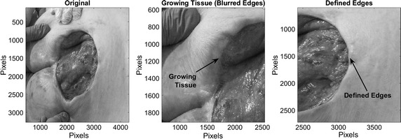
Pressure ulcers have become subject of study in recent years due to the treatment high costs and decreased life quality from patients. These chronic wounds are related to the global life expectancy increment, being the geriatric and physical disable patients the principal affected by this condition. Injuries diagnosis and treatment usually takes weeks or even months by medical personel. Using non-invasive techniques, such as image processing techniques, it is possible to conduct an analysis from ulcers and aid in its diagnosis.

METHODS

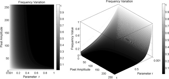
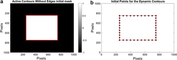
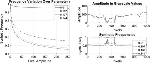
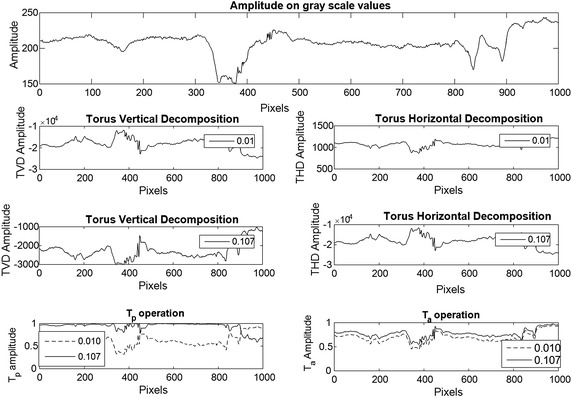
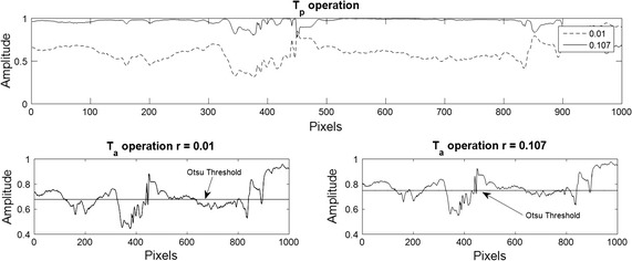
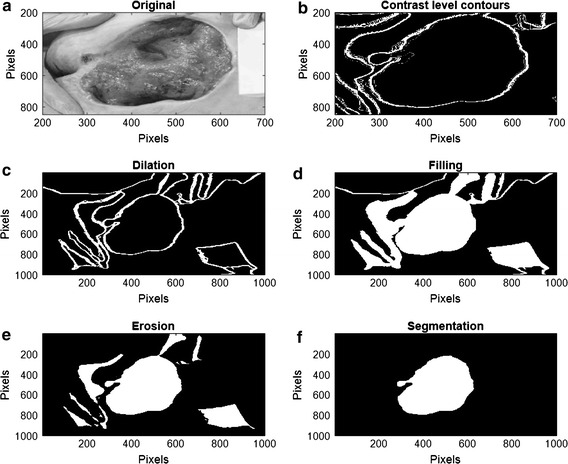
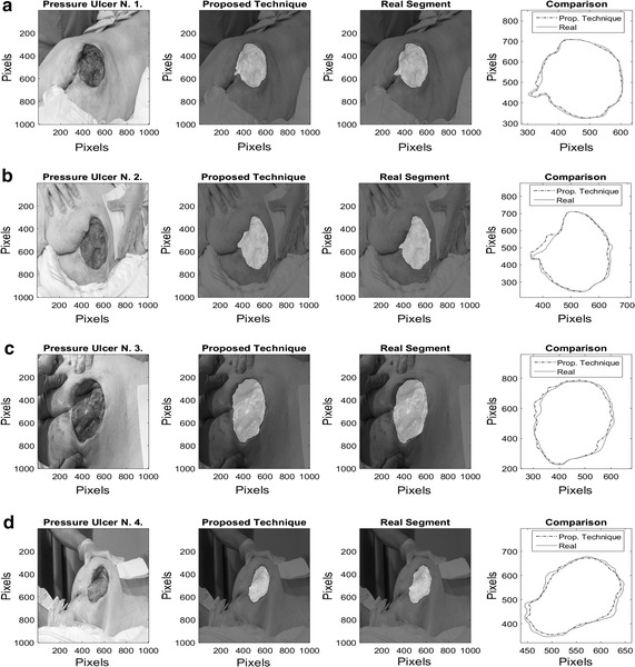
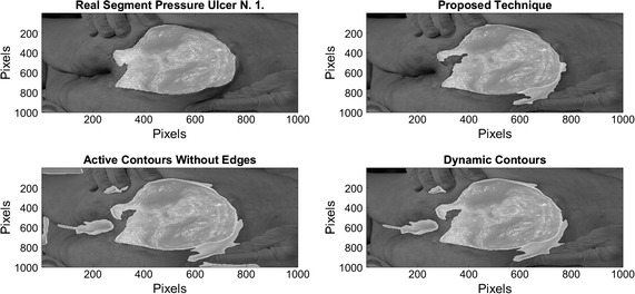
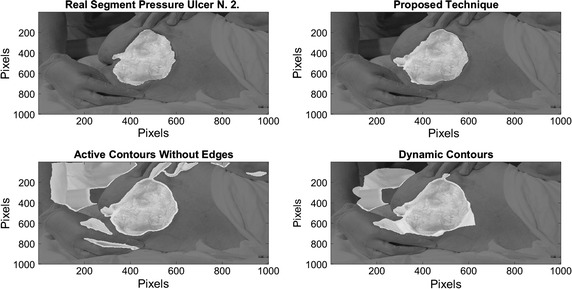
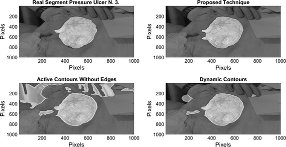
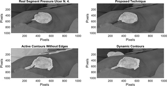
This paper proposes a novel technique for image segmentation based on contrast changes by using synthetic frequencies obtained from the grayscale value available in each pixel of the image. These synthetic frequencies are calculated using the model of energy density over an electric field to describe a relation between a constant density and the image amplitude in a pixel. A toroidal geometry is used to decompose the image into different contrast levels by variating the synthetic frequencies. Then, the decomposed image is binarized applying Otsu's threshold allowing for obtaining the contours that describe the contrast variations. Morphological operations are used to obtain the desired segment of the image.

RESULTS

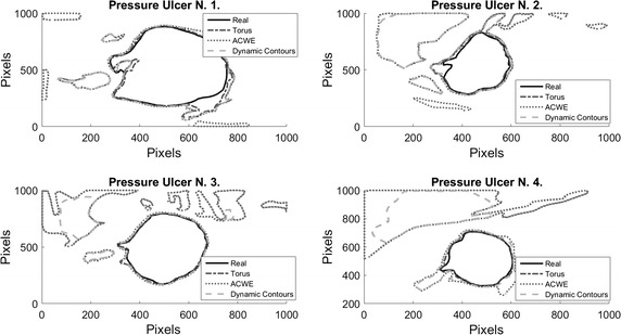
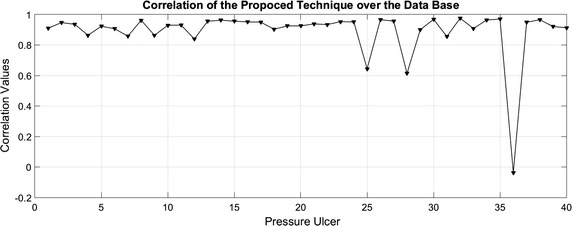
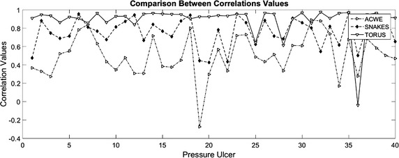
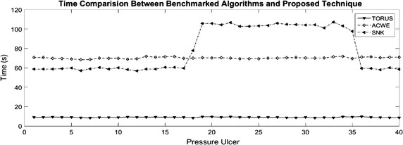
The proposed technique is evaluated by synthesizing a Data Base with 51 images of pressure ulcers, provided by the Centre IGURCO. With the segmentation of these pressure ulcer images it is possible to aid in its diagnosis and treatment. To provide evidences of technique performance, digital image correlation was used as a measure, where the segments obtained using the methodology are compared with the real segments. The proposed technique is compared with two benchmarked algorithms. The results over the technique present an average correlation of 0.89 with a variation of ±0.1 and a computational time of 9.04 seconds.

CONCLUSIONS

The methodology presents better segmentation results than the benchmarked algorithms using less computational time and without the need of an initial condition.

摘要

背景

近年来,由于治疗成本高昂以及患者生活质量下降,压疮已成为研究对象。这些慢性伤口与全球预期寿命的增加有关,老年患者和身体残疾患者是受此状况影响的主要群体。医疗人员对损伤的诊断和治疗通常需要数周甚至数月时间。使用图像处理技术等非侵入性技术,可以对溃疡进行分析并辅助其诊断。

方法

本文提出了一种基于对比度变化的图像分割新技术,该技术利用从图像每个像素的灰度值中获得的合成频率。这些合成频率是使用电场能量密度模型计算得出的,以描述恒定密度与像素中图像幅度之间的关系。采用环形几何结构,通过改变合成频率将图像分解为不同的对比度级别。然后,应用大津阈值对分解后的图像进行二值化处理,从而获得描述对比度变化的轮廓。使用形态学操作来获取图像的所需部分。

结果

通过合成由IGURCO中心提供的包含51张压疮图像的数据库,对所提出的技术进行了评估。通过对这些压疮图像进行分割,可以辅助其诊断和治疗。为了提供技术性能的证据,使用数字图像相关性作为一种度量方法,将使用该方法获得的部分与真实部分进行比较。将所提出的技术与两种基准算法进行了比较。该技术的结果显示平均相关性为0.89,变化范围为±0.1,计算时间为9.04秒。

结论

该方法比基准算法呈现出更好的分割结果,使用的计算时间更少,并且无需初始条件。

https://cdn.ncbi.nlm.nih.gov/pmc/blobs/9877/5234262/6f51759a9692/12938_2016_298_Fig16_HTML.jpg
https://cdn.ncbi.nlm.nih.gov/pmc/blobs/9877/5234262/850bd8a62c24/12938_2016_298_Fig1_HTML.jpg
https://cdn.ncbi.nlm.nih.gov/pmc/blobs/9877/5234262/11ab11027611/12938_2016_298_Fig2_HTML.jpg
https://cdn.ncbi.nlm.nih.gov/pmc/blobs/9877/5234262/1d05d0bf940b/12938_2016_298_Fig3_HTML.jpg
https://cdn.ncbi.nlm.nih.gov/pmc/blobs/9877/5234262/eb832a9462b5/12938_2016_298_Fig4_HTML.jpg
https://cdn.ncbi.nlm.nih.gov/pmc/blobs/9877/5234262/0e9d0af3ccb5/12938_2016_298_Fig5_HTML.jpg
https://cdn.ncbi.nlm.nih.gov/pmc/blobs/9877/5234262/e7a15c951d95/12938_2016_298_Fig6_HTML.jpg
https://cdn.ncbi.nlm.nih.gov/pmc/blobs/9877/5234262/ce4f93f77955/12938_2016_298_Fig7_HTML.jpg
https://cdn.ncbi.nlm.nih.gov/pmc/blobs/9877/5234262/6e07c144ea2c/12938_2016_298_Fig8_HTML.jpg
https://cdn.ncbi.nlm.nih.gov/pmc/blobs/9877/5234262/9d77e798fb4e/12938_2016_298_Fig9_HTML.jpg
https://cdn.ncbi.nlm.nih.gov/pmc/blobs/9877/5234262/dcdee1658c69/12938_2016_298_Fig10_HTML.jpg
https://cdn.ncbi.nlm.nih.gov/pmc/blobs/9877/5234262/37edccd2b1a5/12938_2016_298_Fig11_HTML.jpg
https://cdn.ncbi.nlm.nih.gov/pmc/blobs/9877/5234262/d84b8cbe983f/12938_2016_298_Fig12_HTML.jpg
https://cdn.ncbi.nlm.nih.gov/pmc/blobs/9877/5234262/e01b083b4a18/12938_2016_298_Fig13_HTML.jpg
https://cdn.ncbi.nlm.nih.gov/pmc/blobs/9877/5234262/2ec53e966afb/12938_2016_298_Fig14_HTML.jpg
https://cdn.ncbi.nlm.nih.gov/pmc/blobs/9877/5234262/4787305ef941/12938_2016_298_Fig15_HTML.jpg
https://cdn.ncbi.nlm.nih.gov/pmc/blobs/9877/5234262/6f51759a9692/12938_2016_298_Fig16_HTML.jpg

相似文献

1
Pressure ulcer image segmentation technique through synthetic frequencies generation and contrast variation using toroidal geometry.基于环形几何结构通过合成频率生成和对比度变化的压疮图像分割技术
Biomed Eng Online. 2017 Jan 6;16(1):4. doi: 10.1186/s12938-016-0298-3.
2
Automated framework for accurate segmentation of pressure ulcer images.用于压力性溃疡图像精确分割的自动化框架。
Comput Biol Med. 2017 Nov 1;90:137-145. doi: 10.1016/j.compbiomed.2017.09.015. Epub 2017 Sep 22.
3
Efficient use of mobile devices for quantification of pressure injury images.高效利用移动设备对压力性损伤图像进行量化分析。
Technol Health Care. 2018;26(S1):269-280. doi: 10.3233/THC-174612.
4
Fuzzy spectral clustering for automated delineation of chronic wound region using digital images.基于数字图像的慢性创面区域自动勾画的模糊谱聚类方法
Comput Biol Med. 2017 Oct 1;89:551-560. doi: 10.1016/j.compbiomed.2017.04.004. Epub 2017 Apr 23.
5
Automated measurement of pressure injury through image processing.通过图像处理自动测量压力性损伤。
J Clin Nurs. 2017 Nov;26(21-22):3564-3575. doi: 10.1111/jocn.13726. Epub 2017 Apr 3.
6
A new iterative triclass thresholding technique in image segmentation.图像分割中的一种新的迭代三阈值技术。
IEEE Trans Image Process. 2014 Mar;23(3):1038-46. doi: 10.1109/TIP.2014.2298981.
7
A novel segmentation approach for noisy medical images using intuitionistic fuzzy divergence with neighbourhood-based membership function.一种基于邻域隶属函数的直觉模糊散度的噪声医学图像分割新方法。
J Microsc. 2015 Mar;257(3):187-200. doi: 10.1111/jmi.12200. Epub 2014 Dec 2.
8
Segmentation of MR image using local and global region based geodesic model.基于局部和全局区域的测地线模型对磁共振图像进行分割。
Biomed Eng Online. 2015 Feb 19;14:8. doi: 10.1186/1475-925X-14-8.
9
Robust 2D Otsu's Algorithm for Uneven Illumination Image Segmentation.用于非均匀光照图像分割的鲁棒二维 Otsu 算法。
Comput Intell Neurosci. 2020 Aug 11;2020:5047976. doi: 10.1155/2020/5047976. eCollection 2020.
10
A strategy for blood vessels segmentation based on the threshold which combines statistical and scale space filter. Application to the study of angiogenesis.一种基于阈值的血管分割策略,该阈值结合了统计和尺度空间滤波器。在血管生成研究中的应用。
Comput Methods Programs Biomed. 2006 Apr;82(1):1-9. doi: 10.1016/j.cmpb.2005.10.008. Epub 2006 Mar 23.

引用本文的文献

1
Integrating 3D Model Representation for an Accurate Non-Invasive Assessment of Pressure Injuries with Deep Learning.利用深度学习技术实现三维模型表示,从而对压力性损伤进行准确的非侵入性评估。
Sensors (Basel). 2020 May 21;20(10):2933. doi: 10.3390/s20102933.
2
Detection of Volatile Compounds Emitted by Bacteria in Wounds Using Gas Sensors.利用气体传感器检测伤口中细菌释放的挥发性化合物。
Sensors (Basel). 2019 Mar 28;19(7):1523. doi: 10.3390/s19071523.
3
Classification of pressure ulcer tissues with 3D convolutional neural network.基于 3D 卷积神经网络的压疮组织分类。

本文引用的文献

1
The Burden of Pressure Ulcers in Spain.西班牙压疮的负担
Wounds. 2007 Jul;19(7):201-6.
2
Non-locally regularized segmentation of multiple sclerosis lesion from multi-channel MRI data.基于多通道MRI数据的多发性硬化病变非局部正则化分割
Magn Reson Imaging. 2014 Oct;32(8):1058-66. doi: 10.1016/j.mri.2014.03.006. Epub 2014 Apr 24.
3
Automatic knee cartilage segmentation from multi-contrast MR images using support vector machine classification with spatial dependencies.基于支持向量机分类的多对比度 MR 图像膝关节软骨自动分割方法:空间相关性分析。
Med Biol Eng Comput. 2018 Dec;56(12):2245-2258. doi: 10.1007/s11517-018-1835-y. Epub 2018 Jun 15.
4
Bactericide effect of methylene blue associated with low-level laser therapy in Escherichia coli bacteria isolated from pressure ulcers.亚甲蓝联合低强度激光疗法对从压疮分离出的大肠杆菌的杀菌作用。
Lasers Med Sci. 2018 Nov;33(8):1723-1731. doi: 10.1007/s10103-018-2528-3. Epub 2018 May 9.
5
Efficient use of mobile devices for quantification of pressure injury images.高效利用移动设备对压力性损伤图像进行量化分析。
Technol Health Care. 2018;26(S1):269-280. doi: 10.3233/THC-174612.
Magn Reson Imaging. 2013 Dec;31(10):1731-43. doi: 10.1016/j.mri.2013.06.005. Epub 2013 Jul 15.
4
Distance regularized level set evolution and its application to image segmentation.距离正则化水平集演化及其在图像分割中的应用。
IEEE Trans Image Process. 2010 Dec;19(12):3243-54. doi: 10.1109/TIP.2010.2069690. Epub 2010 Aug 26.
5
Active contours without edges.无边缘活动轮廓。
IEEE Trans Image Process. 2001;10(2):266-77. doi: 10.1109/83.902291.
6
3D ultrasound elastography for early detection of lesions. evaluation on a pressure ulcer mimicking phantom.用于病变早期检测的三维超声弹性成像:在模拟压疮体模上的评估
Annu Int Conf IEEE Eng Med Biol Soc. 2007;2007:79-82. doi: 10.1109/IEMBS.2007.4352227.
7
A non-contact imaging-based approach to detecting stage I pressure ulcers.
Conf Proc IEEE Eng Med Biol Soc. 2006;2006:6380-3. doi: 10.1109/IEMBS.2006.259513.
8
The cost of pressure ulcers in the UK.英国压疮的成本。
Age Ageing. 2004 May;33(3):230-5. doi: 10.1093/ageing/afh086.
9
A review of the etiology and treatment of skin ulcers with wound dressings: comparison of the effects of occlusive and nonocclusive dressings.伤口敷料治疗皮肤溃疡的病因及治疗综述:封闭性敷料与非封闭性敷料效果比较
J Long Term Eff Med Implants. 1992;2(4):267-88.
10
Wound status evaluation using color image processing.使用彩色图像处理技术进行伤口状态评估。
IEEE Trans Med Imaging. 1997 Feb;16(1):78-86. doi: 10.1109/42.552057.