Dawes Timothy J W, de Marvao Antonio, Shi Wenzhe, Fletcher Tristan, Watson Geoffrey M J, Wharton John, Rhodes Christopher J, Howard Luke S G E, Gibbs J Simon R, Rueckert Daniel, Cook Stuart A, Wilkins Martin R, O'Regan Declan P
From the MRC Clinical Sciences Centre, Du Cane Rd, London W12 0NN, England (T.J.W.D., A.d.M., W.S., T.F., S.A.C., D.P.O.); Division of Experimental Medicine, Department of Medicine (T.J.W.D, T.F., G.M.J.W., J.W., C.J.R., M.R.W.), Department of Computing (W.S., D.R.), and National Heart and Lung Institute (J.S.R.G.), Imperial College London, London, England; National Heart Centre Singapore, Singapore and Duke-NUS Graduate Medical School, Singapore (S.A.C.); and Department of Cardiology, National Pulmonary Hypertension Service, Imperial College Healthcare NHS Trust, London, England (L.S.G.E.H., J.S.R.G.).
Radiology. 2017 May;283(2):381-390. doi: 10.1148/radiol.2016161315. Epub 2017 Jan 16.
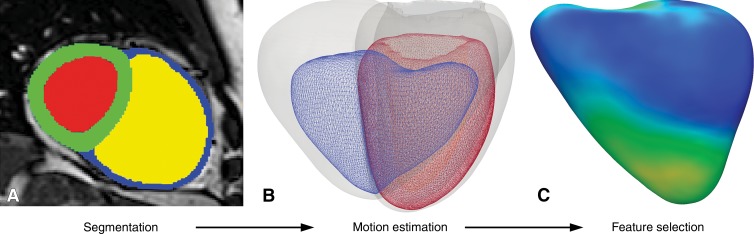
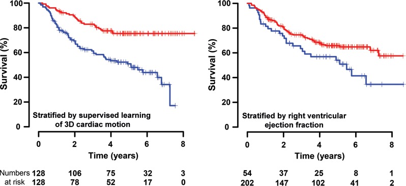
Purpose To determine if patient survival and mechanisms of right ventricular failure in pulmonary hypertension could be predicted by using supervised machine learning of three-dimensional patterns of systolic cardiac motion. Materials and Methods The study was approved by a research ethics committee, and participants gave written informed consent. Two hundred fifty-six patients (143 women; mean age ± standard deviation, 63 years ± 17) with newly diagnosed pulmonary hypertension underwent cardiac magnetic resonance (MR) imaging, right-sided heart catheterization, and 6-minute walk testing with a median follow-up of 4.0 years. Semiautomated segmentation of short-axis cine images was used to create a three-dimensional model of right ventricular motion. Supervised principal components analysis was used to identify patterns of systolic motion that were most strongly predictive of survival. Survival prediction was assessed by using difference in median survival time and area under the curve with time-dependent receiver operating characteristic analysis for 1-year survival. Results At the end of follow-up, 36% of patients (93 of 256) died, and one underwent lung transplantation. Poor outcome was predicted by a loss of effective contraction in the septum and free wall, coupled with reduced basal longitudinal motion. When added to conventional imaging and hemodynamic, functional, and clinical markers, three-dimensional cardiac motion improved survival prediction (area under the receiver operating characteristic curve, 0.73 vs 0.60, respectively; P < .001) and provided greater differentiation according to difference in median survival time between high- and low-risk groups (13.8 vs 10.7 years, respectively; P < .001). Conclusion A machine-learning survival model that uses three-dimensional cardiac motion predicts outcome independent of conventional risk factors in patients with newly diagnosed pulmonary hypertension. Online supplemental material is available for this article.
目的 通过使用收缩期心脏运动三维模式的监督式机器学习来确定肺动脉高压患者的生存情况及右心室衰竭机制是否能够被预测。材料与方法 本研究经研究伦理委员会批准,参与者签署了书面知情同意书。256例新诊断为肺动脉高压的患者(143例女性;平均年龄±标准差,63岁±17岁)接受了心脏磁共振(MR)成像、右心导管检查及6分钟步行试验,中位随访时间为4.0年。使用短轴电影图像的半自动分割创建右心室运动的三维模型。采用监督主成分分析来识别对生存最具预测性的收缩期运动模式。通过中位生存时间差异及1年生存时间依赖型受试者操作特征分析的曲线下面积评估生存预测情况。结果 在随访结束时,36%的患者(256例中的93例)死亡,1例接受了肺移植。室间隔和游离壁有效收缩丧失以及基底纵向运动减少预示着不良预后。当将三维心脏运动添加到传统成像、血流动力学、功能和临床标志物中时,可改善生存预测(受试者操作特征曲线下面积分别为0.73和0.60;P <.001),并根据高风险组和低风险组之间的中位生存时间差异提供更大的区分度(分别为13.8年和10.7年;P <.001)。结论 一种使用三维心脏运动的机器学习生存模型可独立于新诊断肺动脉高压患者的传统风险因素预测预后。本文提供在线补充材料。