Martin Gregory M, Yoshioka Craig, Rex Emily A, Fay Jonathan F, Xie Qing, Whorton Matthew R, Chen James Z, Shyng Show-Ling
Department of Biochemistry and Molecular Biology, Oregon Health and Science University, Portland, Oregon, United States.
Department of Biomedical Engineering, Oregon Health and Science University, Portland, Oregon, United States.
Elife. 2017 Jan 16;6:e24149. doi: 10.7554/eLife.24149.
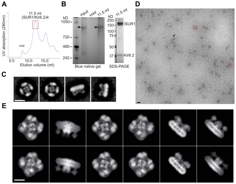
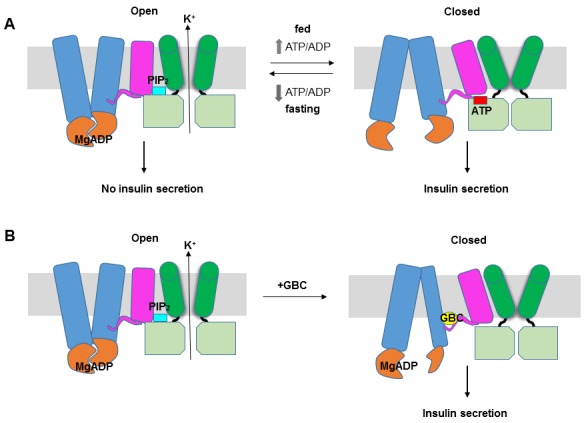
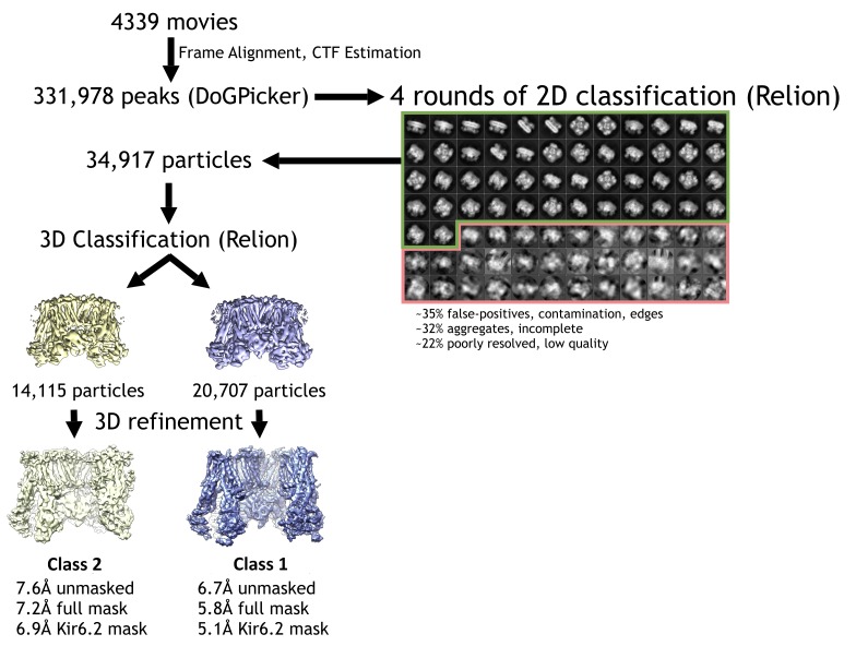
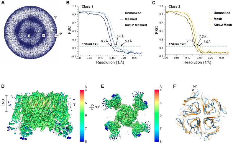
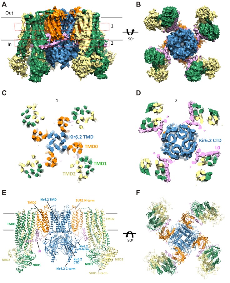
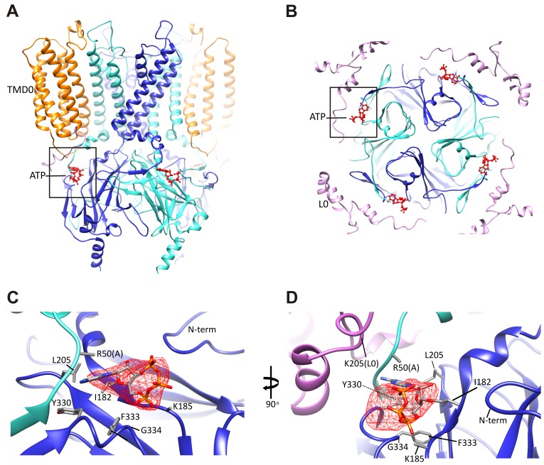
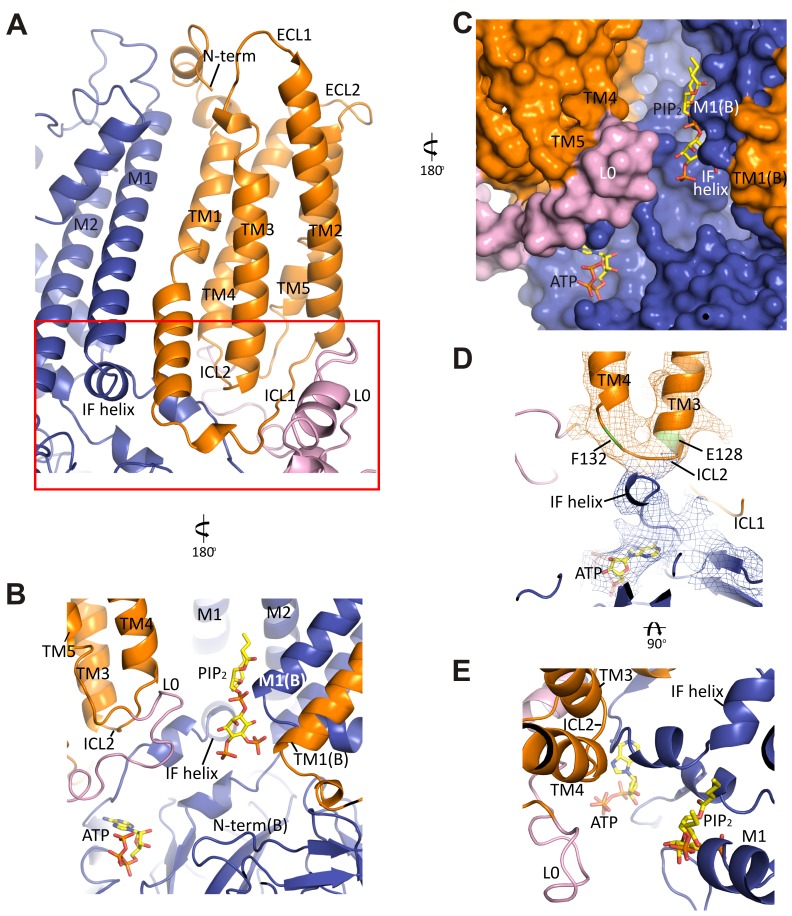
K channels are metabolic sensors that couple cell energetics to membrane excitability. In pancreatic β-cells, channels formed by SUR1 and Kir6.2 regulate insulin secretion and are the targets of antidiabetic sulfonylureas. Here, we used cryo-EM to elucidate structural basis of channel assembly and gating. The structure, determined in the presence of ATP and the sulfonylurea glibenclamide, at ~6 Å resolution reveals a closed Kir6.2 tetrameric core with four peripheral SUR1s each anchored to a Kir6.2 by its N-terminal transmembrane domain (TMD0). Intricate interactions between TMD0, the loop following TMD0, and Kir6.2 near the proposed PIP binding site, and where ATP density is observed, suggest SUR1 may contribute to ATP and PIP binding to enhance Kir6.2 sensitivity to both. The SUR1-ABC core is found in an unusual inward-facing conformation whereby the two nucleotide binding domains are misaligned along a two-fold symmetry axis, revealing a possible mechanism by which glibenclamide inhibits channel activity.
钾通道是将细胞能量代谢与膜兴奋性相耦合的代谢传感器。在胰腺β细胞中,由SUR1和Kir6.2形成的通道调节胰岛素分泌,并且是抗糖尿病磺脲类药物的作用靶点。在这里,我们使用冷冻电镜来阐明通道组装和门控的结构基础。在存在ATP和磺脲类药物格列本脲的情况下,以约6 Å分辨率测定的结构揭示了一个封闭的Kir6.2四聚体核心,其周围有四个SUR1,每个SUR1通过其N端跨膜结构域(TMD0)锚定到一个Kir6.2上。TMD0、TMD0之后的环以及Kir6.2在拟议的磷脂酰肌醇(PIP)结合位点附近且观察到ATP密度的位置之间的复杂相互作用表明,SUR1可能有助于ATP和PIP结合,以增强Kir6.2对两者的敏感性。发现SUR1-ABC核心处于一种不寻常的向内构象,其中两个核苷酸结合结构域沿二重对称轴未对齐,揭示了格列本脲抑制通道活性的一种可能机制。