Erchova Irina, Vasalauskaite Asta, Longo Valentina, Sengpiel Frank
School of Biosciences and Neuroscience and Mental Health Research Institute, Cardiff University, Sir Martin Evans Building, Museum Avenue, Cardiff, CF10 3AX, UK.
School of Biosciences and Neuroscience and Mental Health Research Institute, Cardiff University, Sir Martin Evans Building, Museum Avenue, Cardiff, CF10 3AX, UK
Philos Trans R Soc Lond B Biol Sci. 2017 Mar 5;372(1715). doi: 10.1098/rstb.2016.0159.
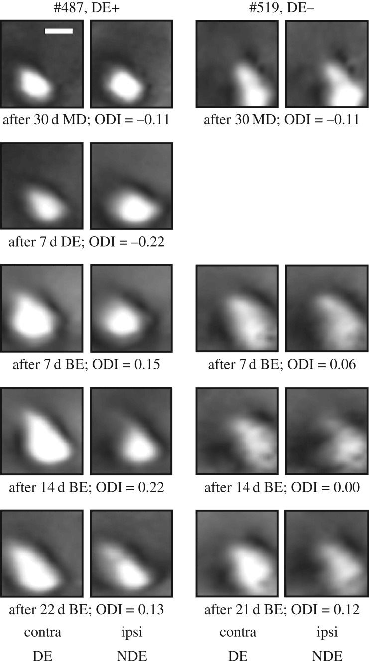
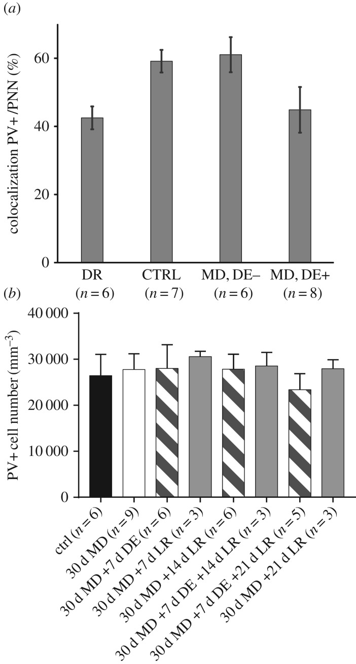
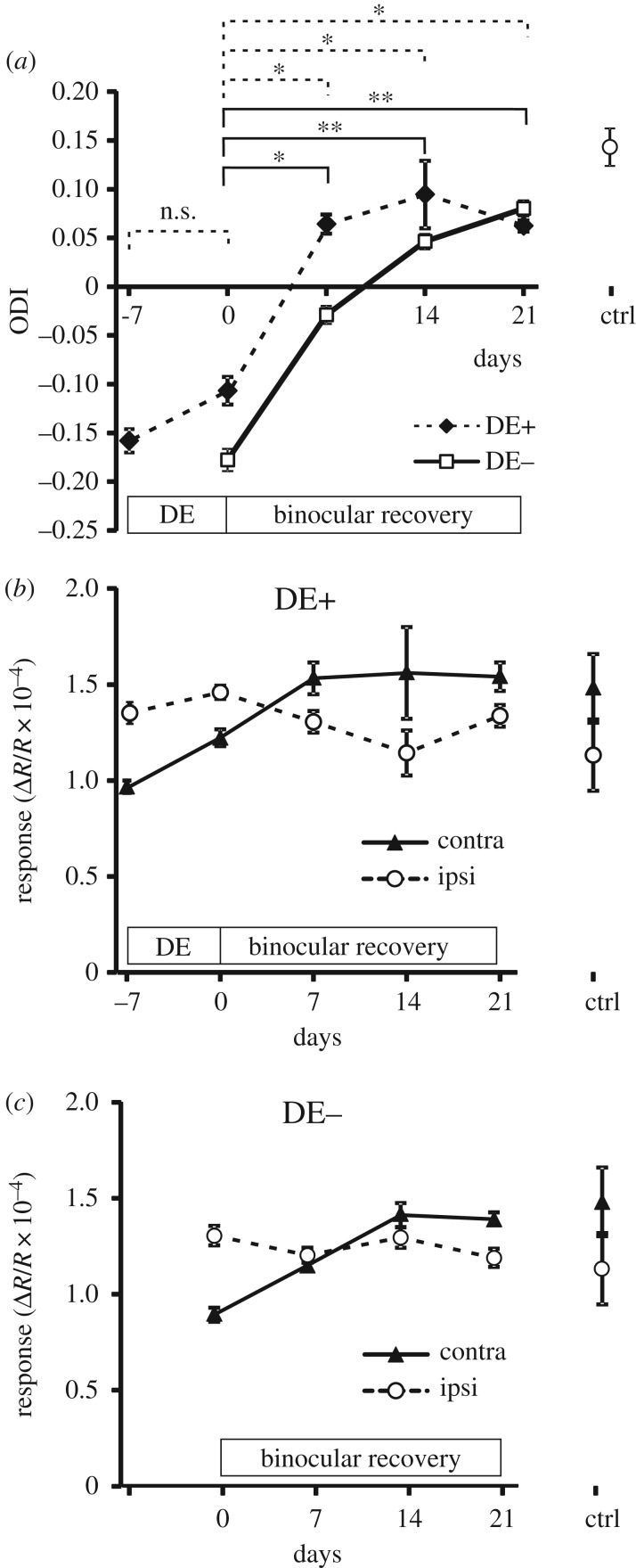
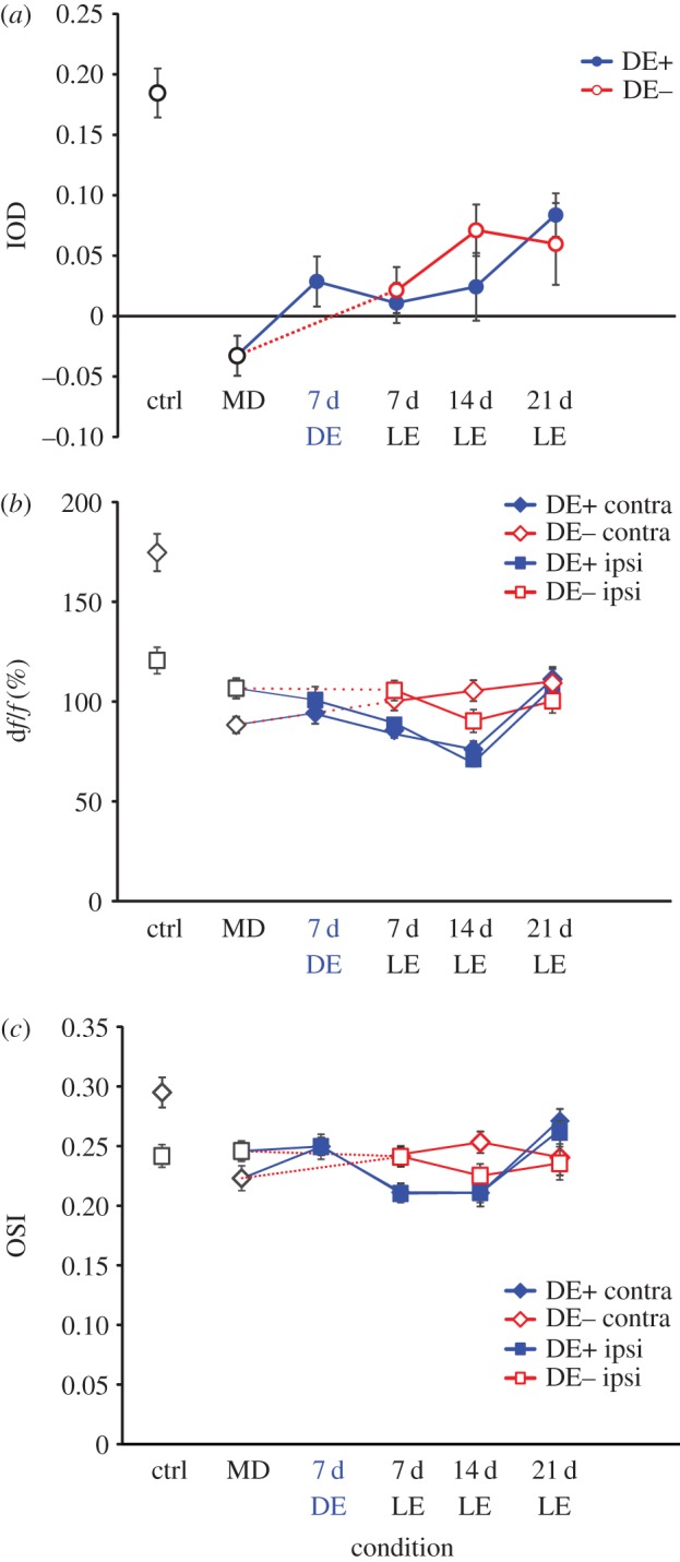
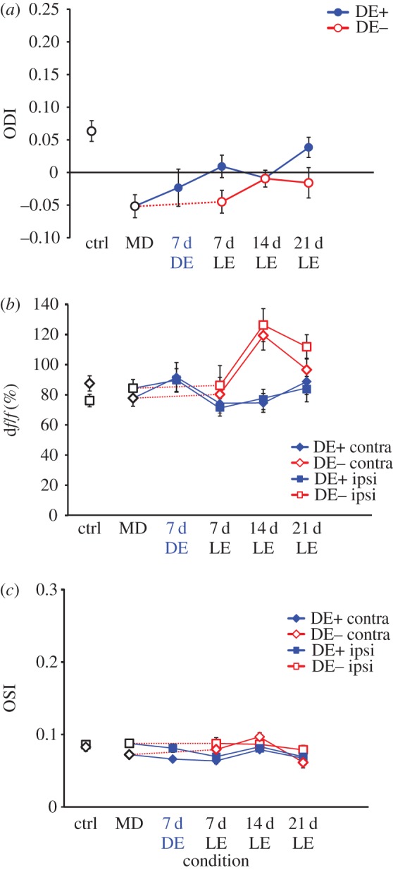
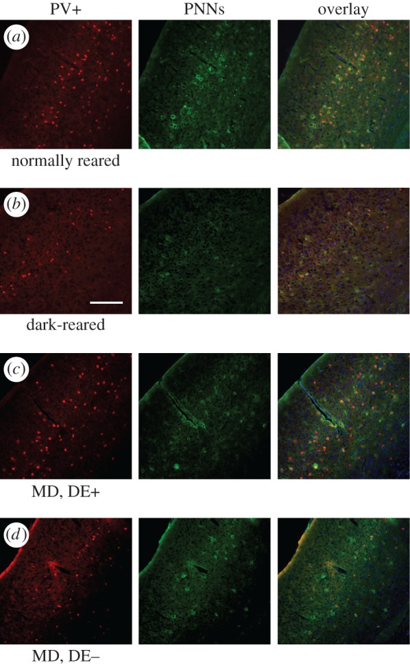
Dark rearing is known to delay the time course of the critical period for ocular dominance plasticity in the visual cortex. Recent evidence suggests that a period of dark exposure (DE) may enhance or reinstate plasticity even after closure of the critical period, mediated through modification of the excitatory-inhibitory balance and/or removal of structural brakes on plasticity. Here, we investigated the effects of a week of DE on the recovery from a month of monocular deprivation (MD) in the primary visual cortex (V1) of juvenile mice. Optical imaging of intrinsic signals revealed that ocular dominance in V1 of mice that had received DE recovered slightly more quickly than of mice that had not, but the level of recovery after three weeks was similar in both groups. Two-photon calcium imaging showed no significant difference in the recovery of orientation selectivity of excitatory neurons between the two groups. Parvalbumin-positive (PV+) interneurons exhibited a smaller ocular dominance shift during MD but again no differences in subsequent recovery. The percentage of PV+ cells surrounded by perineuronal nets, a structural brake on plasticity, was lower in mice with than those without DE. Overall, DE causes a modest enhancement of mouse visual cortex plasticity.This article is part of the themed issue 'Integrating Hebbian and homeostatic plasticity'.
已知暗饲养会延迟视觉皮层中眼优势可塑性关键期的时间进程。最近的证据表明,即使在关键期关闭后,一段时间的暗暴露(DE)也可能通过改变兴奋性-抑制性平衡和/或消除可塑性的结构限制来增强或恢复可塑性。在此,我们研究了一周的DE对幼年小鼠初级视觉皮层(V1)中一个月单眼剥夺(MD)恢复的影响。内在信号的光学成像显示,接受DE的小鼠V1中的眼优势恢复速度比未接受DE的小鼠略快,但三周后的恢复水平在两组中相似。双光子钙成像显示,两组兴奋性神经元方向选择性的恢复没有显著差异。小白蛋白阳性(PV+)中间神经元在MD期间表现出较小的眼优势偏移,但随后的恢复也没有差异。被神经元周围网包围的PV+细胞百分比(可塑性的一种结构限制),在接受DE的小鼠中比未接受DE的小鼠更低。总体而言,DE会适度增强小鼠视觉皮层的可塑性。本文是主题为“整合赫布可塑性和稳态可塑性”特刊的一部分。