Oliveira Rodrigo Ribeiro de, Medina de Mattos Rômulo, Magalhães Rebelo Luciana, Guimarães Meireles Ferreira Fernanda, Tovar-Moll Fernanda, Eurico Nasciutti Luiz, de Castro Brito Gerly Anne
Inter-institutional Doctorate Program in Morphological Science, Federal University of Ceará / Federal University of Rio de Janeiro, Rio de Janeiro, Brazil.
Department of Physical Therapy, Federal University of Ceara, Fortaleza, Ceará, Brazil.
PLoS One. 2017 Jan 17;12(1):e0169513. doi: 10.1371/journal.pone.0169513. eCollection 2017.
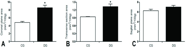
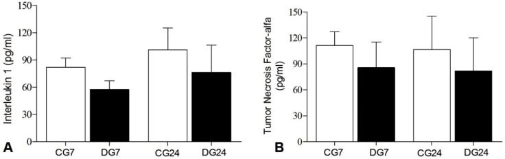
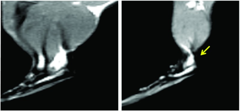
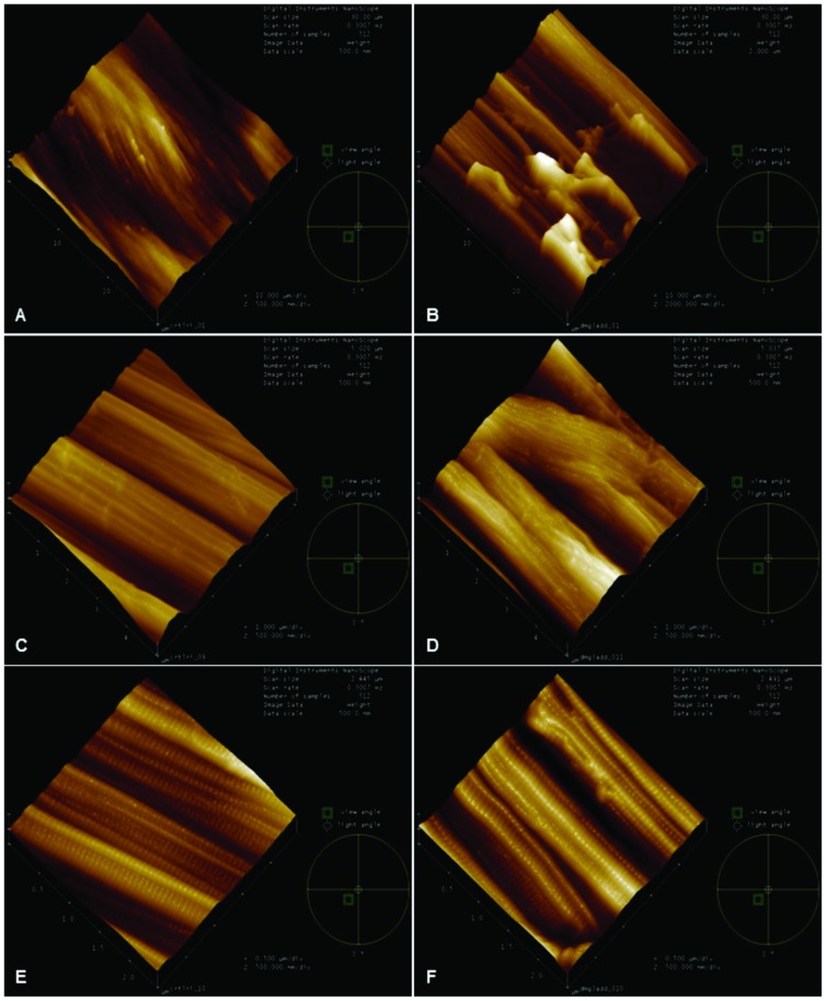
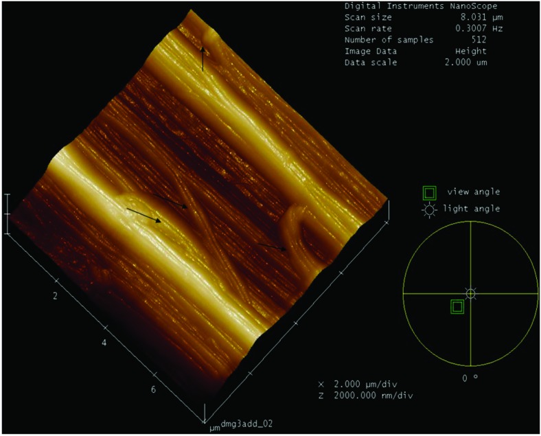
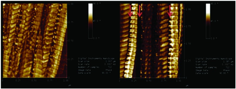
Although of several studies that associate chronic hyperglycemia with tendinopathy, the connection between morphometric changes as witnessed by magnetic resonance (MR) images, nanostructural changes, and inflammatory markers have not yet been fully established. Therefore, the present study has as a hypothesis that the Achilles tendons of rats with diabetes mellitus (DM) exhibit structural changes. The animals were randomly divided into two experimental groups: Control Group (n = 06) injected with a vehicle (sodium citrate buffer solution) and Diabetic Group (n = 06) consisting of rats submitted to intraperitoneal administration of streptozotocin. MR was performed 24 days after the induction of diabetes and images were used for morphometry using ImageJ software. Morphology of the collagen fibers within tendons was examined using Atomic Force microscopy (AFM). An increase in the dimension of the coronal plane area was observed in the diabetic group (8.583 ± 0.646 mm2/100g) when compared to the control group (4.823 ± 0.267 mm2/100g) resulting in a significant difference (p = 0.003) upon evaluating the Achilles tendons. Similarly, our analysis found an increase in the size of the transverse section area in the diabetic group (1.328 ± 0.103 mm2/100g) in comparison to the control group (0.940 ± 0.01 mm2/100g) p = 0.021. The tendons of the diabetic group showed great irregularity in fiber bundles, including modified grain direction and jagged junctions and deformities in the form of collagen fibrils bulges. Despite the morphological changes observed in the Achilles tendon of diabetic animals, IL1 and TNF-α did not change. Our results suggest that DM promotes changes to the Achilles tendon with important structural modifications as seen by MR and AFM, excluding major inflammatory changes.
尽管有多项研究将慢性高血糖与肌腱病联系起来,但磁共振(MR)图像所显示的形态计量学变化、纳米结构变化和炎症标志物之间的联系尚未完全确立。因此,本研究提出一个假设,即糖尿病(DM)大鼠的跟腱会出现结构变化。动物被随机分为两个实验组:对照组(n = 06)注射赋形剂(柠檬酸钠缓冲溶液),糖尿病组(n = 06)由腹腔注射链脲佐菌素的大鼠组成。在糖尿病诱导后24天进行磁共振成像,并使用ImageJ软件对图像进行形态计量分析。使用原子力显微镜(AFM)检查肌腱内胶原纤维的形态。与对照组(4.823±0.267mm²/100g)相比,糖尿病组(8.583±0.646mm²/100g)的冠状面面积尺寸增加,在评估跟腱时差异具有统计学意义(p = 0.003)。同样,我们的分析发现,与对照组(0.940±0.01mm²/100g)相比,糖尿病组的横截面积尺寸增加(1.328±0.103mm²/100g),p = 0.021。糖尿病组的肌腱在纤维束方面表现出很大的不规则性,包括纹理方向改变、锯齿状连接以及胶原纤维凸起形式的畸形。尽管在糖尿病动物的跟腱中观察到了形态学变化,但白细胞介素1和肿瘤坏死因子-α并未改变。我们的结果表明,糖尿病会促使跟腱发生变化,磁共振成像和原子力显微镜观察到其具有重要的结构改变,但不包括主要的炎症变化。