Qin Huamin, Janowski Miroslaw, Pearl Monica S, Malysz-Cymborska Izabela, Li Shen, Eberhart Charles G, Walczak Piotr
Russell H. Morgan Department. of Radiology and Radiological Science, Division of MR Research, Cellular Imaging Section, The Johns Hopkins University School of Medicine, Baltimore, MD, United States of America.
Institute for Cell Engineering, The Johns Hopkins University School of Medicine, Baltimore, MD, United States of America.
PLoS One. 2017 Jan 19;12(1):e0169656. doi: 10.1371/journal.pone.0169656. eCollection 2017.
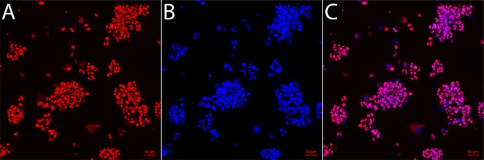
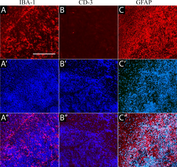
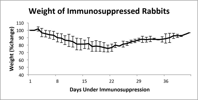
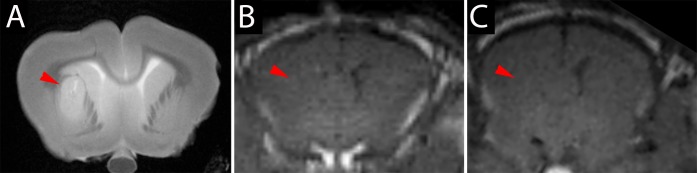
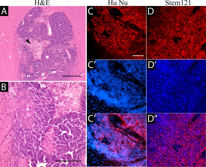
The prognosis for malignant brain tumors remains poor despite a combination of surgery, radiotherapy, and chemotherapy. This is partly due to the blood-brain barrier, a major obstacle that prevents therapeutic agents from effectively reaching the tumor. We have recently developed a method for precise and predictable opening of the blood-brain barrier via the intra-arterial administration of mannitol, a hyperosmolar agent, in a rabbit model, whose vascular anatomy facilitates the use of standard interventional neuroradiology techniques and devices. To date, however, no protocols are available that enable human glioma modeling in rabbits. In this article, we report on the xenotransplantation of a human glioblastoma (GBM-1) in adult New Zealand rabbits. We induced multi-drug immunosuppression (Mycophenolate Mofetil, Dexamethasone, Tacrolimus) and stereotactically implanted GBM-1 tumor cells into rabbit brains. The rabbits were followed for 42 days, monitored by MRI and body weight measurements, and underwent postmortem histopathological analysis. On MRI, brain tumors were identified on T2-weighted scans. On histopathology, tumors were detected with hematoxylin/eosin and their human origin was confirmed with immunohistochemistry against human-specific antigens. Our method for human glioma modeling in rabbits provides the foundation to test novel treatment strategies, including intra-arterial therapeutic agent delivery.
尽管采用了手术、放疗和化疗相结合的方法,恶性脑肿瘤的预后仍然很差。部分原因是血脑屏障,这是一个主要障碍,阻止治疗药物有效到达肿瘤部位。我们最近在兔模型中开发了一种通过动脉内注射甘露醇(一种高渗剂)精确且可预测地打开血脑屏障的方法,兔的血管解剖结构便于使用标准的介入神经放射学技术和设备。然而,迄今为止,尚无能够在兔体内建立人胶质瘤模型的方案。在本文中,我们报告了在成年新西兰兔体内异种移植人胶质母细胞瘤(GBM - 1)的情况。我们诱导多药免疫抑制(霉酚酸酯、地塞米松、他克莫司),并将GBM - 1肿瘤细胞立体定向植入兔脑。对兔进行了42天的随访,通过MRI和体重测量进行监测,并进行死后组织病理学分析。在MRI的T2加权扫描上发现了脑肿瘤。在组织病理学上,用苏木精/伊红检测到肿瘤,并用针对人类特异性抗原的免疫组织化学证实了其人类起源。我们在兔体内建立人胶质瘤模型的方法为测试包括动脉内给药在内的新型治疗策略奠定了基础。