Division of Thoracic Surgery, Department of Surgery, Toronto General Hospital, University of Toronto, University Health Network, Toronto, Canada.
PLoS One. 2013 Jun 28;8(6):e67355. doi: 10.1371/journal.pone.0067355. Print 2013.
The rabbit VX2 lung cancer model is a large animal model useful for preclinical lung cancer imaging and interventional studies. However, previously reported models had issues in terms of invasiveness of tumor inoculation, control of tumor aggressiveness and incidence of complications.
We aimed to develop a minimally invasive rabbit VX2 lung cancer model suitable for imaging and transbronchial interventional studies.
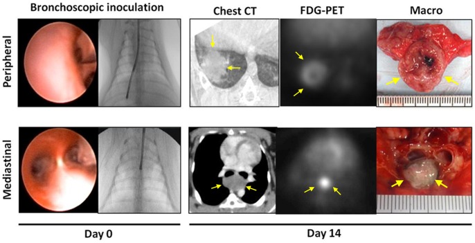
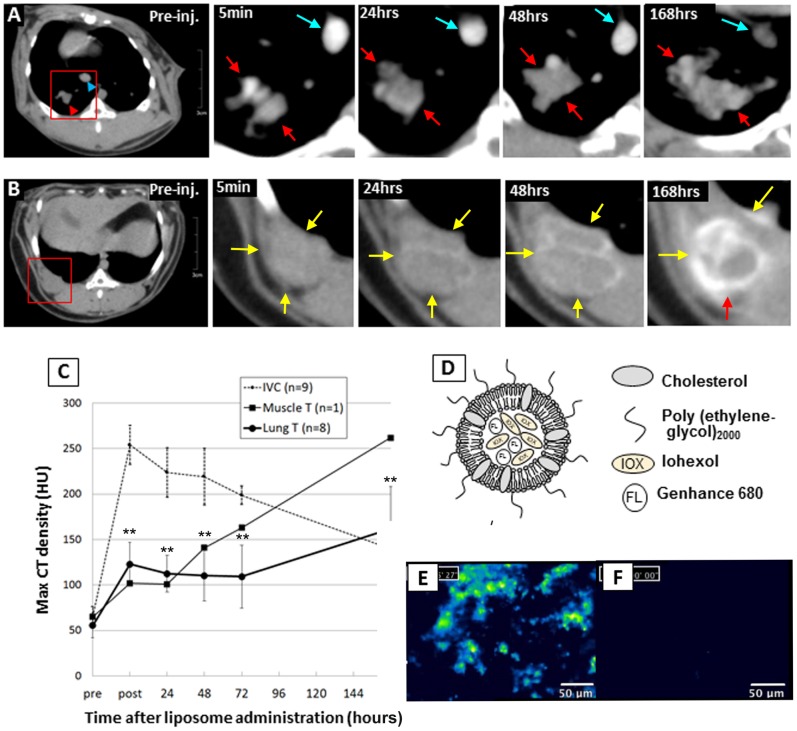
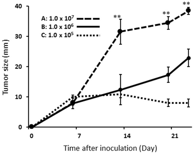
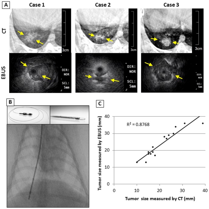
New Zealand white rabbits and VX2 tumors were used in the study. An ultra-thin bronchoscope was inserted through a miniature laryngeal mask airway into the bronchus. Different numbers of VX2 tumor cells were selectively inoculated into the lung parenchyma or subcarinal mediastinum to create a uniform tumor with low incidence of complications. The model was characterized by CT, FDG-PET, and endobronchial ultrasound (EBUS). Liposomal dual-modality contrast agent was used to evaluate liposome drug delivery system in this model.
Both peripheral and mediastinal lung tumor models were created. The tumor making success rate was 75.8% (25/33) in the peripheral lung tumor model and 60% (3/5) in the mediastinal tumor model. The group of 1.0×10(6) of VX2 tumor cells inoculation showed a linear growth curve with less incidence of complications. Radial probe EBUS visualized the internal structure of the tumor and the size measurement correlated well with CT measurements (r(2) = 0.98). Over 7 days of continuous enhancement of the lung tumor by liposomal contrast in the lung tumor was confirmed both CT and fluorescence imaging.
Our minimally invasive bronchoscopic rabbit VX2 lung cancer model is an ideal platform for lung cancer imaging and preclinical bronchoscopic interventional studies.
兔 VX2 肺癌模型是一种用于临床前肺癌成像和介入研究的大型动物模型。然而,以前报道的模型在肿瘤接种的侵袭性、肿瘤侵袭性的控制和并发症的发生率方面存在问题。
我们旨在开发一种适用于成像和经支气管介入研究的微创兔 VX2 肺癌模型。
本研究使用新西兰白兔和 VX2 肿瘤。将超薄支气管镜通过微型喉罩气道插入支气管。将不同数量的 VX2 肿瘤细胞选择性接种到肺实质或隆突下纵隔,以创建一种具有低并发症发生率的均匀肿瘤。该模型通过 CT、FDG-PET 和支气管内超声(EBUS)进行了特征描述。使用脂质体双模态造影剂评估了该模型中的脂质体药物传递系统。
成功构建了外周和纵隔肺肿瘤模型。在外周肺肿瘤模型中,肿瘤制作成功率为 75.8%(25/33),在纵隔肿瘤模型中为 60%(3/5)。接种 1.0×10(6)个 VX2 肿瘤细胞的组显示出线性生长曲线,并发症发生率较低。径向探头 EBUS 可视化了肿瘤的内部结构,并且大小测量与 CT 测量相关性良好(r(2) = 0.98)。在肺癌中,脂质体造影剂连续增强 7 天,在 CT 和荧光成像中均证实了肺肿瘤的增强。
我们的微创支气管镜兔 VX2 肺癌模型是一种理想的肺癌成像和临床前支气管镜介入研究平台。