Vasquez Kristine O, Peterson Jeffrey D
PerkinElmer Inc., Hopkinton, Massachusetts.
PerkinElmer Inc., Hopkinton, Massachusetts
J Pharmacol Exp Ther. 2017 Apr;361(1):87-98. doi: 10.1124/jpet.116.238378. Epub 2017 Jan 23.
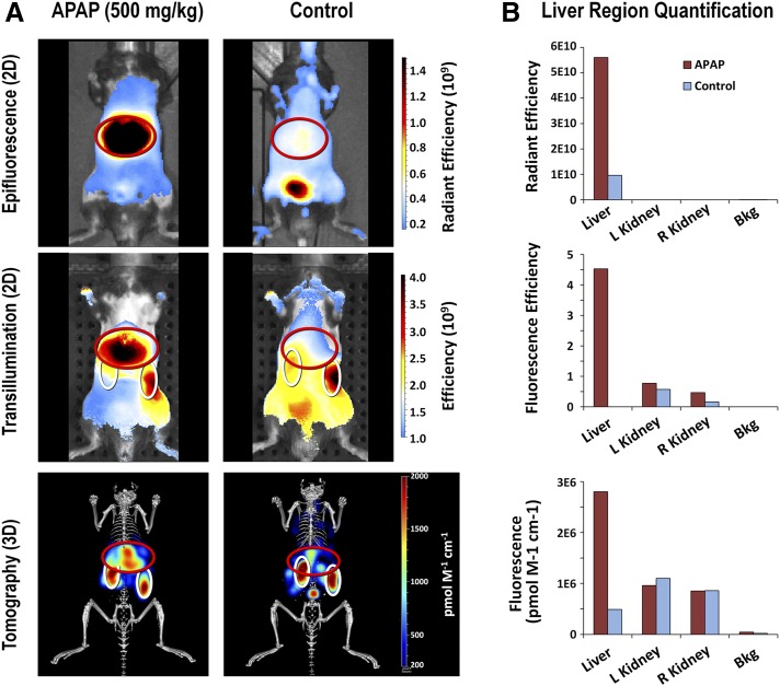
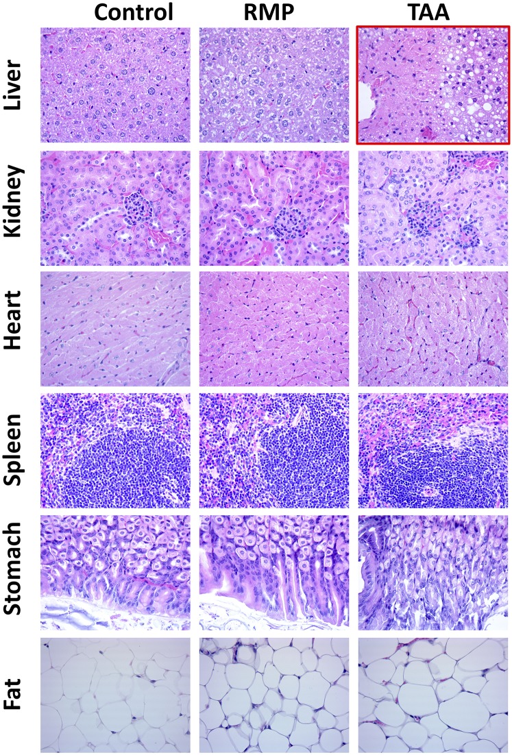
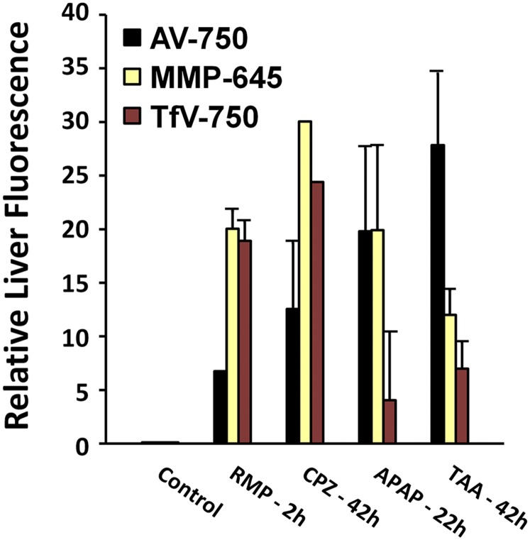
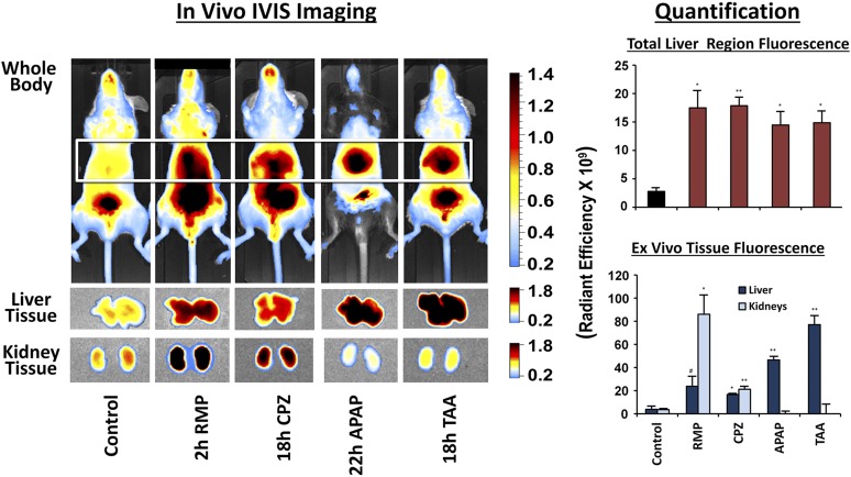
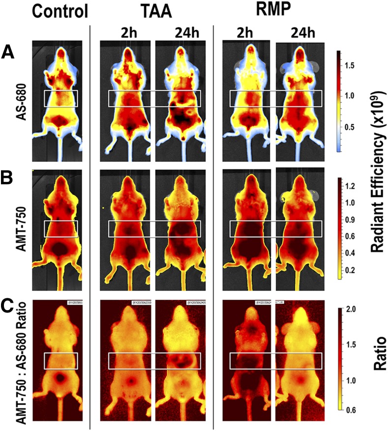
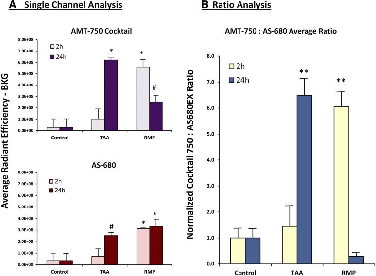
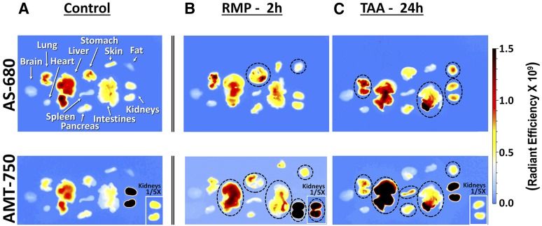
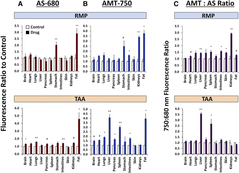
Hepatocellular and cholestatic forms of drug-induced liver injury (DILI) are major reasons for late-stage termination of small-molecule drug discovery research projects. Biochemical serum markers are limited in their ability to sensitively and specifically detect both of these common DILI forms in preclinical models, and tissue-specific approaches to assessing this are labor intensive, requiring extensive animal dosing, tissue preparation, and pathology assessment. In vivo fluorescent imaging offers noninvasive detection of biologic changes detected directly in the livers of living animals. Three different near-infrared fluorescent imaging probes, specific for cell death (Annexin-Vivo 750), matrix metalloproteases (MMPSense 750 FAST), and transferrin receptor (Transferrin-Vivo 750) were used to measure the effects of single bolus intraperitoneal doses of four different chemical agents known to induce liver injury. Hepatocellular injury-inducing agents, thioacetamide and acetaminophen, showed optimal injury detection with probe injection at 18-24 hours, the liver cholestasis-inducing drug rifampicin required early probe injection (2 hours), and chlorpromazine, which induces mixed hepatocellular/cholestatic injury, showed injury with both early and late injection. Different patterns of liver responses were seen among these different imaging probes, and no one probe detected injury by all four compounds. By using a cocktail of these three near-infrared fluorescent imaging probes, all labeled with 750-nm fluorophores, each of the four different DILI agents induced comparable tissue injury within the liver region, as assessed by epifluorescence imaging. A strategy of probe cocktail injection in separate cohorts at 2 hours and at 20-24 hours allowed the effective detection of drugs with either early- or late-onset injury.
肝细胞型和胆汁淤积型药物性肝损伤(DILI)是小分子药物发现研究项目后期终止的主要原因。生化血清标志物在临床前模型中灵敏且特异检测这两种常见DILI类型的能力有限,而评估此情况的组织特异性方法劳动强度大,需要大量动物给药、组织制备和病理学评估。体内荧光成像可对活体动物肝脏中直接检测到的生物学变化进行无创检测。使用三种不同的近红外荧光成像探针,分别针对细胞死亡(膜联蛋白 - 体内750)、基质金属蛋白酶(基质金属蛋白酶传感750 FAST)和转铁蛋白受体(转铁蛋白 - 体内750),来测量腹腔单次推注四种已知可诱导肝损伤的不同化学试剂的效果。诱导肝细胞损伤的试剂硫代乙酰胺和对乙酰氨基酚,在注射探针后18 - 24小时显示出最佳损伤检测效果,诱导肝胆汁淤积的药物利福平需要早期注射探针(2小时),而诱导混合性肝细胞/胆汁淤积性损伤的氯丙嗪,早期和晚期注射均显示有损伤。在这些不同的成像探针中观察到不同的肝脏反应模式,没有一种探针能检测到所有四种化合物造成的损伤。通过使用这三种均标记有750纳米荧光团的近红外荧光成像探针混合物,通过落射荧光成像评估,四种不同的DILI试剂在肝脏区域均诱导了相当的组织损伤。在不同组中分别于2小时和20 - 24小时注射探针混合物的策略,能够有效检测具有早期或晚期损伤的药物。