• 文献检索
  • 文档翻译
  • 深度研究
  • 学术资讯
  • Suppr Zotero 插件Zotero 插件
  • 邀请有礼
  • 套餐&价格
  • 历史记录
应用&插件
Suppr Zotero 插件Zotero 插件浏览器插件Mac 客户端Windows 客户端微信小程序
定价
高级版会员购买积分包购买API积分包
服务
文献检索文档翻译深度研究API 文档MCP 服务
关于我们
关于 Suppr公司介绍联系我们用户协议隐私条款
关注我们

Suppr 超能文献

核心技术专利:CN118964589B侵权必究
粤ICP备2023148730 号-1Suppr @ 2026

文献检索

告别复杂PubMed语法,用中文像聊天一样搜索,搜遍4000万医学文献。AI智能推荐,让科研检索更轻松。

立即免费搜索

文件翻译

保留排版,准确专业,支持PDF/Word/PPT等文件格式,支持 12+语言互译。

免费翻译文档

深度研究

AI帮你快速写综述,25分钟生成高质量综述,智能提取关键信息,辅助科研写作。

立即免费体验

鉴定在心室肌细胞死亡后存在于缺血人心肌中的心脏祖细胞。

Identification of cardiac progenitors that survive in the ischemic human heart after ventricular myocyte death.

机构信息

Department of Physiology, Shiga University of Medical Science, Otsu, Shiga, 520-2192, Japan.

Department of Pathology, Shiga University of Medical Science, Otsu, Shiga, 520-2192, Japan.

出版信息

Sci Rep. 2017 Jan 25;7:41318. doi: 10.1038/srep41318.

DOI:10.1038/srep41318
PMID:28120944
原文链接:https://pmc.ncbi.nlm.nih.gov/articles/PMC5264617/
Abstract

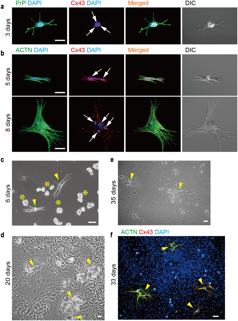
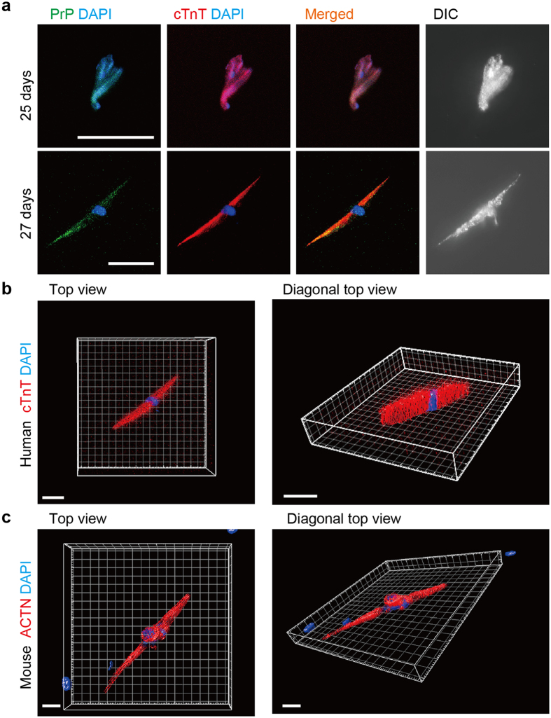
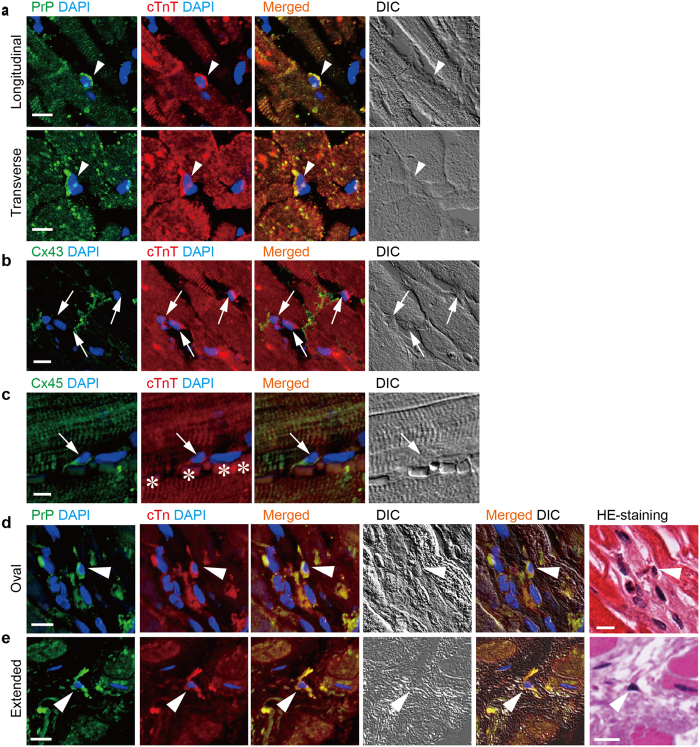
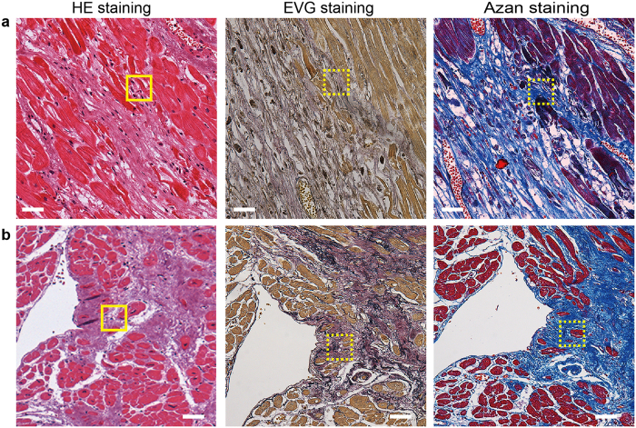
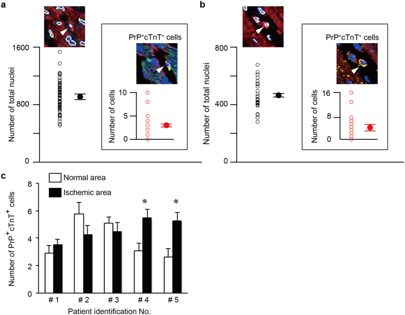
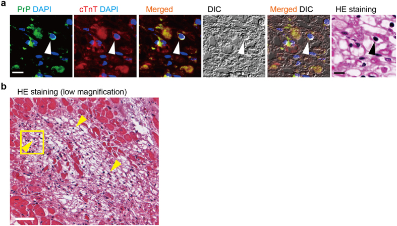
Atypically-shaped cardiomyocytes (ACMs) are beating heart cells identified in the cultures of cardiomyocyte-removed fractions obtained from adult mouse hearts. Since ACMs spontaneously develop into beating cells in the absence of hormones or chemicals, these cells are likely to be a type of cardiac progenitors rather than stem cells. "Native ACMs" are found as small interstitial cells among ventricular myocytes that co-express cellular prion protein (PrP) and cardiac troponin T (cTnT) in mouse and human heart tissues. However, the endogenous behavior of human ACMs is unclear. In the present study, we demonstrate that PrP cTnT cells are present in the human heart tissue with myocardial infarction (MI). These cells were mainly found in the border of necrotic cardiomyocytes caused by infarcts and also in the hibernating myocardium subjected to the chronic ischemia. The ratio of PrP cTnT cells to the total cells observed in the normal heart tissue section of mouse and human was estimated to range from 0.3-0.8%. Notably, living human PrP cTnT cells were identified in the cultures obtained at pathological autopsy despite exposure to lethal ischemic conditions for hours after death. These findings suggest that ACMs could survive in the ischemic human heart and develop into a sub-population of cardiac myocytes.

摘要

非典型形态心肌细胞(ACMs)是在从成年小鼠心脏中去除心肌细胞的培养物中鉴定出的搏动心脏细胞。由于 ACMs 在没有激素或化学物质的情况下自发发育为搏动细胞,因此这些细胞可能是一种心脏祖细胞,而不是干细胞。“天然 ACMs”作为小的间质细胞存在于心室肌细胞之间,在小鼠和人心脏组织中共同表达细胞朊病毒蛋白(PrP)和心肌肌钙蛋白 T(cTnT)。然而,人 ACMs 的内源性行为尚不清楚。在本研究中,我们证明了 PrP cTnT 细胞存在于心肌梗死(MI)的人类心脏组织中。这些细胞主要存在于梗死引起的坏死心肌细胞的边界处,也存在于慢性缺血引起的冬眠心肌中。在小鼠和人正常心脏组织切片中观察到的 PrP cTnT 细胞与总细胞的比例估计在 0.3-0.8%之间。值得注意的是,尽管在死亡后数小时内暴露于致命的缺血条件下,仍在尸检时获得的培养物中鉴定出存活的人类 PrP cTnT 细胞。这些发现表明,ACMs 可以在缺血的人心肌中存活并发育成心肌细胞的亚群。

https://cdn.ncbi.nlm.nih.gov/pmc/blobs/971a/5264617/660dfaf01615/srep41318-f6.jpg
https://cdn.ncbi.nlm.nih.gov/pmc/blobs/971a/5264617/b6bc3af5447b/srep41318-f1.jpg
https://cdn.ncbi.nlm.nih.gov/pmc/blobs/971a/5264617/109142be006e/srep41318-f2.jpg
https://cdn.ncbi.nlm.nih.gov/pmc/blobs/971a/5264617/abbfbb549280/srep41318-f3.jpg
https://cdn.ncbi.nlm.nih.gov/pmc/blobs/971a/5264617/84ba97ce769b/srep41318-f4.jpg
https://cdn.ncbi.nlm.nih.gov/pmc/blobs/971a/5264617/42f68154264a/srep41318-f5.jpg
https://cdn.ncbi.nlm.nih.gov/pmc/blobs/971a/5264617/660dfaf01615/srep41318-f6.jpg

相似文献

1
Identification of cardiac progenitors that survive in the ischemic human heart after ventricular myocyte death.鉴定在心室肌细胞死亡后存在于缺血人心肌中的心脏祖细胞。
Sci Rep. 2017 Jan 25;7:41318. doi: 10.1038/srep41318.
2
Prion protein- and cardiac troponin T-marked interstitial cells from the adult myocardium spontaneously develop into beating cardiomyocytes.来自成年心肌的朊病毒蛋白和心肌肌钙蛋白T标记的间质细胞可自发发育成跳动的心肌细胞。
Sci Rep. 2014 Dec 3;4:7301. doi: 10.1038/srep07301.
3
Ischemic survival and constitutively active autophagy in self-beating atypically-shaped cardiomyocytes (ACMs): characterization of a new subpopulation of heart cells.自搏性非典型心肌细胞(ACMs)中的缺血存活和组成性活跃自噬:一种新型心脏细胞亚群的特征。
J Physiol Sci. 2013 Jan;63(1):17-29. doi: 10.1007/s12576-012-0236-5. Epub 2012 Oct 6.
4
Atypically Shaped Cardiomyocytes (ACMs): The Identification, Characterization and New Insights into a Subpopulation of Cardiomyocytes.形态异常的心肌细胞(ACMs):一种心肌细胞亚群的鉴定、特征描述和新认识。
Biomolecules. 2022 Jun 27;12(7):896. doi: 10.3390/biom12070896.
5
Dedifferentiation, Proliferation, and Redifferentiation of Adult Mammalian Cardiomyocytes After Ischemic Injury.成年哺乳动物心肌细胞在缺血性损伤后的去分化、增殖和再分化
Circulation. 2017 Aug 29;136(9):834-848. doi: 10.1161/CIRCULATIONAHA.116.024307. Epub 2017 Jun 22.
6
Cardiac troponin in ischemic cardiomyocytes: intracellular decrease before onset of cell death.缺血性心肌细胞中心肌肌钙蛋白:细胞死亡前的细胞内减少。
Exp Mol Pathol. 2014 Jun;96(3):339-45. doi: 10.1016/j.yexmp.2014.02.012. Epub 2014 Mar 4.
7
Higher frequencies of BCRP+ cardiac resident cells in ischaemic human myocardium.缺血性人类心肌中 BCRP+ 心脏驻留细胞的频率更高。
Eur Heart J. 2013 Sep;34(36):2830-8. doi: 10.1093/eurheartj/ehs156. Epub 2012 Jun 26.
8
A novel type of self-beating cardiomyocytes in adult mouse ventricles.成年小鼠心室中一种新型的自搏动心肌细胞。
Biochem Biophys Res Commun. 2009 Apr 10;381(3):361-6. doi: 10.1016/j.bbrc.2009.02.048. Epub 2009 Feb 15.
9
Structural remodelling of cardiomyocytes in the border zone of infarcted rabbit heart.梗死兔心脏边缘区心肌细胞的结构重塑
Mol Cell Biochem. 2007 Aug;302(1-2):225-32. doi: 10.1007/s11010-007-9445-2. Epub 2007 Mar 27.
10
C-terminal variable AGES domain of Thymosin β4: the molecule's primary contribution in support of post-ischemic cardiac function and repair.四肽素β 4 的 C 末端可变 AGES 结构域:该分子对支持缺血后心脏功能和修复的主要贡献。
J Mol Cell Cardiol. 2015 Oct;87:113-25. doi: 10.1016/j.yjmcc.2015.07.004. Epub 2015 Aug 5.

引用本文的文献

1
Atypically Shaped Cardiomyocytes (ACMs): The Identification, Characterization and New Insights into a Subpopulation of Cardiomyocytes.形态异常的心肌细胞(ACMs):一种心肌细胞亚群的鉴定、特征描述和新认识。
Biomolecules. 2022 Jun 27;12(7):896. doi: 10.3390/biom12070896.
2
Simultaneous Isolation and Culture of Atrial Myocytes, Ventricular Myocytes, and Non-Myocytes from an Adult Mouse Heart.从成年小鼠心脏中同时分离和培养心房肌细胞、心室肌细胞和非肌细胞。
J Vis Exp. 2020 Jun 14(160). doi: 10.3791/61224.
3
Inhibition of Senescence-Associated Genes Rb1 and Meis2 in Adult Cardiomyocytes Results in Cell Cycle Reentry and Cardiac Repair Post-Myocardial Infarction.

本文引用的文献

1
Effect of Bone Marrow-Derived Mononuclear Cell Treatment, Early or Late After Acute Myocardial Infarction: Twelve Months CMR and Long-Term Clinical Results.骨髓单个核细胞治疗对急性心肌梗死后早期或晚期的影响:12 个月的心脏磁共振和长期临床结果。
Circ Res. 2016 Jul 22;119(3):481-90. doi: 10.1161/CIRCRESAHA.116.308639. Epub 2016 Jun 6.
2
Hypoxia fate mapping identifies cycling cardiomyocytes in the adult heart.低氧命运图谱鉴定成年心脏中的循环心肌细胞。
Nature. 2015 Jul 9;523(7559):226-30. doi: 10.1038/nature14582. Epub 2015 Jun 22.
3
Origin of cardiomyocytes in the adult heart.
抑制衰老相关基因 Rb1 和 Meis2 可使成年心肌细胞重新进入细胞周期,并在心肌梗死后修复心脏。
J Am Heart Assoc. 2019 Aug 6;8(15):e012089. doi: 10.1161/JAHA.119.012089. Epub 2019 Jul 18.
4
A simple antegrade perfusion method for isolating viable single cardiomyocytes from neonatal to aged mice.一种从新生小鼠到老年小鼠分离存活单个心肌细胞的简单顺行灌注方法。
Physiol Rep. 2018 May;6(9):e13688. doi: 10.14814/phy2.13688.
5
Use of DAVID algorithms for clustering custom annotated gene lists in a non-model organism, rainbow trout.使用DAVID算法对非模式生物虹鳟鱼中自定义注释的基因列表进行聚类。
BMC Res Notes. 2018 Jan 23;11(1):63. doi: 10.1186/s13104-018-3154-7.
成年心脏中心肌细胞的起源。
Circ Res. 2015 Jan 2;116(1):150-66. doi: 10.1161/CIRCRESAHA.116.303595.
4
Prion protein- and cardiac troponin T-marked interstitial cells from the adult myocardium spontaneously develop into beating cardiomyocytes.来自成年心肌的朊病毒蛋白和心肌肌钙蛋白T标记的间质细胞可自发发育成跳动的心肌细胞。
Sci Rep. 2014 Dec 3;4:7301. doi: 10.1038/srep07301.
5
Existing cardiomyocytes generate cardiomyocytes at a low rate after birth in mice.出生后,在小鼠中,现有的心肌细胞以低速率产生心肌细胞。
Proc Natl Acad Sci U S A. 2014 Jun 17;111(24):8850-5. doi: 10.1073/pnas.1408233111. Epub 2014 May 29.
6
Cardiac stem cell therapy and the promise of heart regeneration.心脏干细胞治疗与心脏再生的前景。
Cell Stem Cell. 2013 Jun 6;12(6):689-98. doi: 10.1016/j.stem.2013.05.008.
7
Cardiomyocyte proliferation and progenitor cell recruitment underlie therapeutic regeneration after myocardial infarction in the adult mouse heart.心肌细胞增殖和祖细胞募集是成年鼠心肌梗死后治疗性再生的基础。
EMBO Mol Med. 2013 Feb;5(2):191-209. doi: 10.1002/emmm.201201737. Epub 2013 Jan 29.
8
Mammalian heart renewal by pre-existing cardiomyocytes.哺乳动物的心肌再生由预先存在的心肌细胞完成。
Nature. 2013 Jan 17;493(7432):433-6. doi: 10.1038/nature11682. Epub 2012 Dec 5.
9
Cardiac progenitors derived from reprogrammed mesenchymal stem cells contribute to angiomyogenic repair of the infarcted heart.重编程间充质干细胞来源的心脏祖细胞有助于梗死心脏的血管生成修复。
Basic Res Cardiol. 2012 Nov;107(6):301. doi: 10.1007/s00395-012-0301-5. Epub 2012 Oct 18.
10
Ischemic survival and constitutively active autophagy in self-beating atypically-shaped cardiomyocytes (ACMs): characterization of a new subpopulation of heart cells.自搏性非典型心肌细胞(ACMs)中的缺血存活和组成性活跃自噬:一种新型心脏细胞亚群的特征。
J Physiol Sci. 2013 Jan;63(1):17-29. doi: 10.1007/s12576-012-0236-5. Epub 2012 Oct 6.