Onoda Atsuto, Takeda Ken, Umezawa Masakazu
Department of Hygienic Chemistry, Graduate School of Pharmaceutical Sciences, Tokyo University of Science, 2641 Yamazaki, Noda, Chiba, 278-8510, Japan.
The Center for Environmental Health Science for the Next Generation, Research Institute for Science and Technology, Organization for Research Advancement, Tokyo University of Science, 2641 Yamazaki, Noda, Chiba, 278-8510, Japan.
Part Fibre Toxicol. 2017 Feb 2;14(1):4. doi: 10.1186/s12989-017-0184-6.
Recent studies indicate that maternal exposure to ambient ultrafine particles and nanoparticles has adverse effects of on the central nervous system. Quantitative dose-response data is required to better understand the developmental neurotoxicity of nanoparticles. The present study investigated dose-dependent effects of maternal exposure to carbon black nanoparticle (CB-NP) on astrocyte in the brains of mouse offspring.
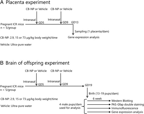
A CB-NP suspension (2.9, 15, or 73 μg/kg) was intranasally administered to pregnant ICR mice on gestational days 5 and 9. Cerebral cortex samples were collected from 6-week-old offspring and examined by Western blotting, immunostaining, microarray analysis, and quantitative reverse transcriptase-polymerase chain reaction. Placentae were collected from pregnant dams on gestational day 13 and examined by microarray analysis.
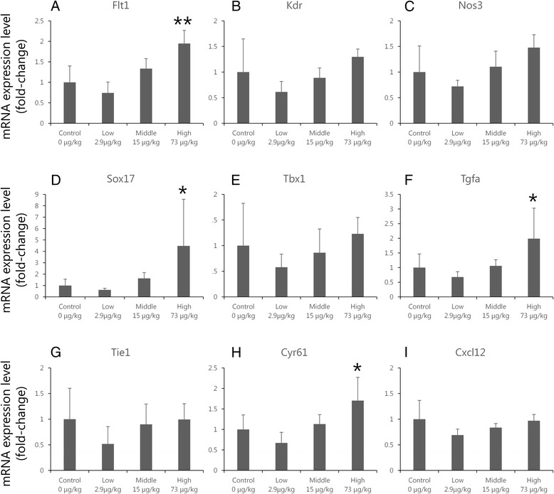
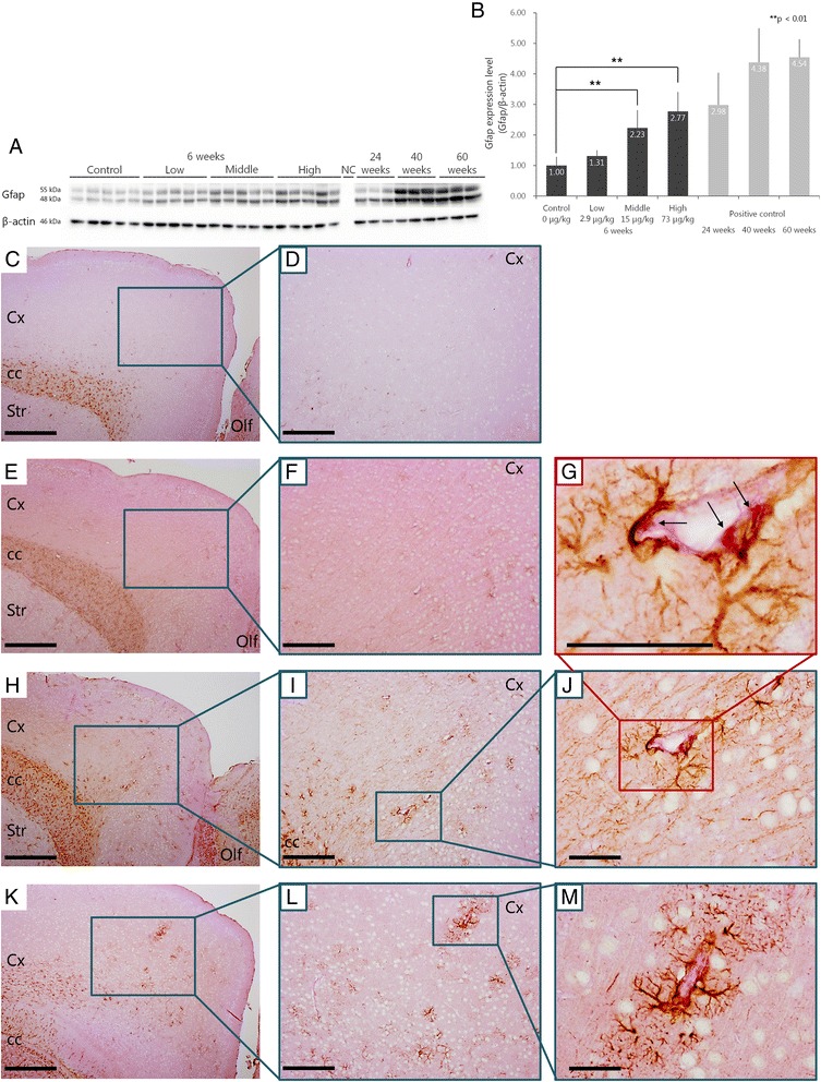
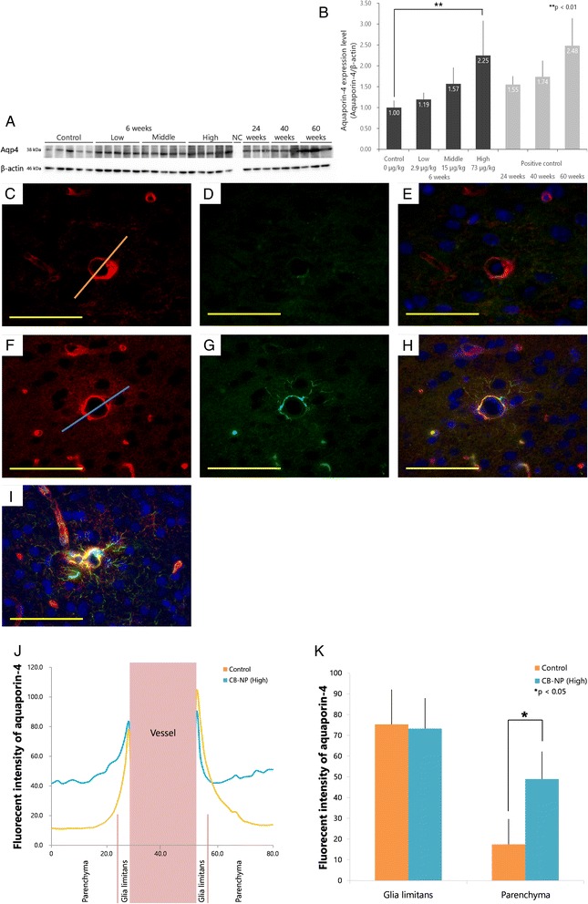
Maternal exposure to CB-NP induced a dose-dependent increase in glial fibrillary acidic protein (GFAP) expression in the cerebral cortex; this increase was particularly observed in astrocytic end-feet attached to denatured perivascular macrophages. Moreover, maternal CB-NP exposure dose-dependently increased aquaporin-4 expression in the brain parenchyma region around blood vessels. The changes in the expression profiles of GFAP and Aqp4 in offspring after maternal CB-NP exposure were similar to those observed in mice of a more advanced age. The expression levels of mRNAs associated with angiogenesis, cell migration, proliferation, chemotaxis, and growth factor production were also altered in the cerebral cortex of offspring after maternal CB-NP exposure. Differentially expressed genes in placental tissues after CB-NP exposure did not populate any specific gene ontology category.
Maternal CB-NP exposure induced long-term activation of astrocytes resulting in reactive astrogliosis in the brains of young mice. Our observations suggest a potentially increased risk of the onset of age-related neurodegenerative diseases by maternal NP exposure. In this study, we report for the first time a quantitative dose-response relationship between maternal NP exposure and phenotypic changes in the central nervous system of the offspring. Moreover, our findings indicate that cortical GFAP and Aqp4 are useful biomarkers that can be employed in further studies aiming to elucidate the underlying mechanism of nanoparticle-mediated developmental neurotoxicity.
近期研究表明,母体暴露于环境超细颗粒和纳米颗粒会对中枢神经系统产生不良影响。需要定量剂量反应数据来更好地了解纳米颗粒的发育神经毒性。本研究调查了母体暴露于炭黑纳米颗粒(CB-NP)对小鼠后代大脑中星形胶质细胞的剂量依赖性影响。
在妊娠第5天和第9天,将CB-NP悬浮液(2.9、15或73μg/kg)经鼻给予怀孕的ICR小鼠。收集6周龄后代的大脑皮质样本,通过蛋白质免疫印迹法、免疫染色、基因芯片分析和定量逆转录聚合酶链反应进行检测。在妊娠第13天从怀孕的母鼠收集胎盘,并通过基因芯片分析进行检测。
母体暴露于CB-NP会导致大脑皮质中胶质纤维酸性蛋白(GFAP)表达呈剂量依赖性增加;这种增加在附着于变性血管周围巨噬细胞的星形胶质细胞终足中尤为明显。此外,母体暴露于CB-NP会使血管周围脑实质区域的水通道蛋白4表达呈剂量依赖性增加。母体暴露于CB-NP后,后代中GFAP和Aqp4表达谱的变化与年龄较大的小鼠中观察到的变化相似。母体暴露于CB-NP后,后代大脑皮质中与血管生成、细胞迁移、增殖、趋化性和生长因子产生相关的mRNA表达水平也发生了改变。CB-NP暴露后胎盘组织中差异表达的基因未归入任何特定的基因本体类别。
母体暴露于CB-NP会导致星形胶质细胞长期激活,从而在幼鼠大脑中引发反应性星形胶质细胞增生。我们的观察结果表明,母体暴露于纳米颗粒可能会增加与年龄相关的神经退行性疾病发病的风险。在本研究中首次报道了母体暴露于纳米颗粒与后代中枢神经系统表型变化之间的定量剂量反应关系。此外,我们的研究结果表明,皮质GFAP和Aqp4是有用的生物标志物,可用于进一步研究以阐明纳米颗粒介导的发育神经毒性的潜在机制。