Shetty Heeresh, Sontakke Subodh, Karjodkar Freny, Gupta Pankaj, Mandwe Ashish, Banga K S
MDS, Assistant Professor, Department of Conservative Dentistry and Endodontics, Nair Hospital Dental College, Mumbai, India.
MDS, Associate Professor, Department of Dentistry Grant Medical College and Sir JJ Group of Hospitals, Mumbai, India.
J Clin Exp Dent. 2017 Jan 1;9(1):e51-e55. doi: 10.4317/jced.52716. eCollection 2017 Jan.
Current technological advances have allowed application of different study designs and techniques for investigation of dental anatomy. Some clinical studies have provided evidence that Cone Beam computed tomography (CBCT) scanning is an important resource in assessment of root canal systems notably to identify MB2 canals in maxillary molars as CBCT scans allow dental investigation in axial, sagittal and coronal planes simultaneously. The current study was undertaken to detect and evaluate filled/unfilled MB2 canals in endodontically treated, asymptomatic maxillary molars utilizing cone beam computed tomography (CBCT).
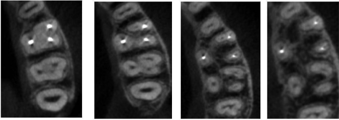
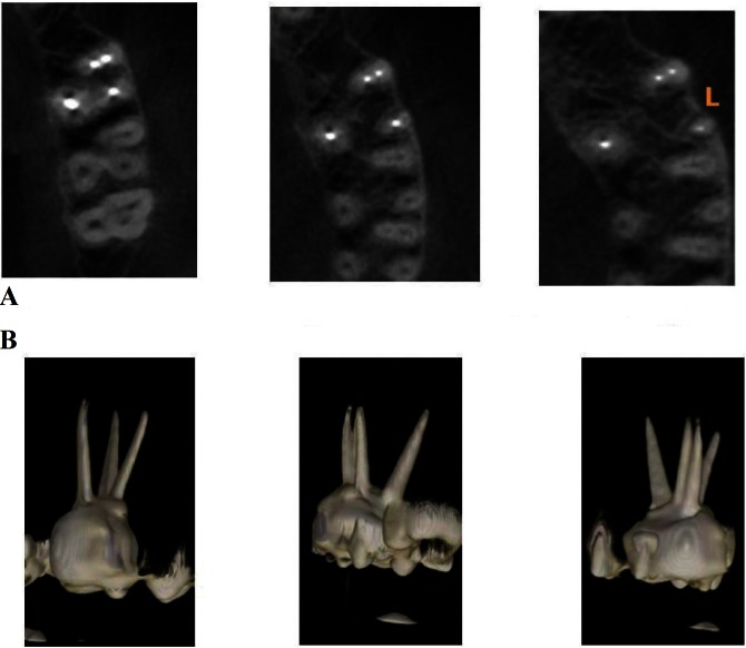
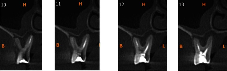
A retrospective study of 100 CBCTs of patients were underwent scanning for various treatment modalities, with asymptomatic endodontically treated permanent first and second maxillary molars were selected. History of root canal treatment varied from minimum of 1 year to a maximum of 10 years. Axial and paraxial images obtained were used to assess the presence of MB2 canal. Paraxial images were used to assess the periapical status.
Of the 100 scans, 66 were of permanent maxillary first molar and 34 were of permanent maxillary second molar. The incidence of MB2 canal was 86.36% in maxillary first molars and 29.4% in maxillary second molars. 77.19 % of maxillary first molars and 90% of maxillary second molars had an unfilled MB2 canal. 72.7% of maxillary first molars and 88.8% of maxillary second molars showed significant periapical radiolucencies in unfilled MB2 canals.
MB2 canals were present in majority of cases and most of the unfilled MB2 canals showed evidence of periapical radiolucencies. MB2 Canals, Cone Beam computed Tomography (CBCT), Filled /Unfilled canals, Endodontically treated teeth.
当前的技术进步使得不同的研究设计和技术可应用于牙体解剖学研究。一些临床研究已提供证据表明,锥形束计算机断层扫描(CBCT)是评估根管系统的重要手段,尤其是在识别上颌磨牙的MB2根管方面,因为CBCT扫描能够同时在轴向、矢状面和冠状面进行牙齿检查。本研究旨在利用锥形束计算机断层扫描(CBCT)检测并评估经根管治疗且无症状的上颌磨牙中已充填/未充填的MB2根管。
对100例接受过各种治疗方式扫描的患者的CBCT进行回顾性研究,选择经根管治疗且无症状的上颌第一和第二恒磨牙。根管治疗史最短1年,最长10年。所获得的轴向和近轴图像用于评估MB2根管的存在情况。近轴图像用于评估根尖周状况。
100次扫描中,66次为上颌第一恒磨牙,34次为上颌第二恒磨牙。上颌第一磨牙MB2根管的发生率为86.36%,上颌第二磨牙为29.4%。上颌第一磨牙77.19%、上颌第二磨牙90%存在未充填的MB2根管。上颌第一磨牙72.7%、上颌第二磨牙88.8%的未充填MB2根管显示有明显的根尖周透射区。
大多数病例中存在MB2根管,且大多数未充填的MB2根管显示有根尖周透射区的迹象。MB2根管、锥形束计算机断层扫描(CBCT)、已充填/未充填根管、经根管治疗的牙齿。