Gorka Stephanie M, Lieberman Lynne, Shankman Stewart A, Phan K Luan
Department of Psychiatry, University of Illinois at Chicago, Chicago, Illinois, USA.
Department of Psychology, University of Illinois at Chicago, Chicago, Illinois, USA.
Psychophysiology. 2017 May;54(5):652-662. doi: 10.1111/psyp.12829. Epub 2017 Feb 2.
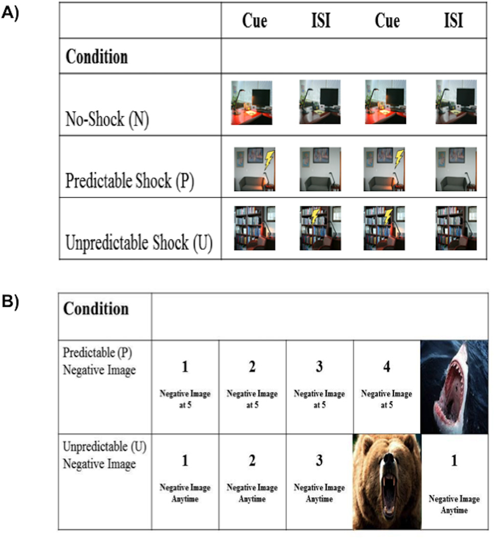
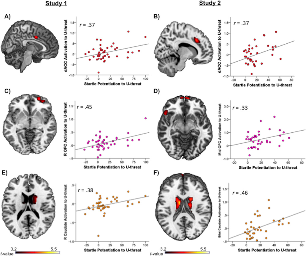
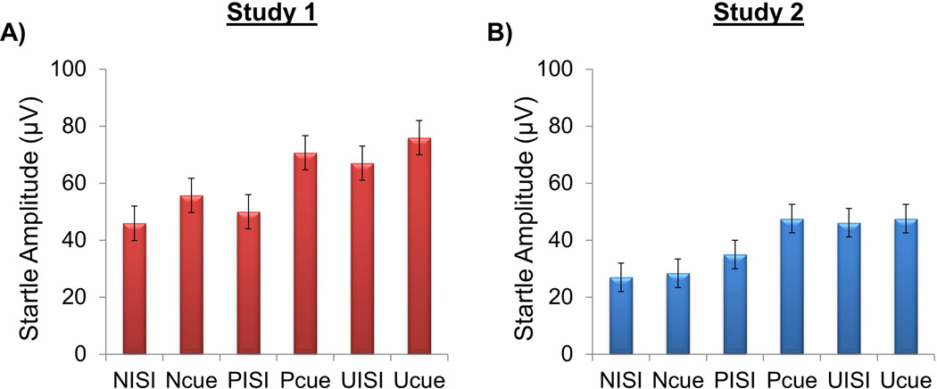
Prior studies indicate that anxiety disorders are associated with heightened sensitivity to uncertain threat (U threat). Individual differences in reactivity to U threat have been measured in the laboratory with two methodologies-startle eyeblink potentiation and fMRI. While startle and fMRI are purported to relate to each other, very little research exists on whether individual differences in one measure are associated with individual differences in another and, thus, whether startle and fMRI capture shared mechanisms. Therefore, the current study was designed to investigate if and where in the brain measures of startle potentiation and fMRI BOLD signal correlate during response to U threat across two independent samples. Participants in both studies completed two threat anticipation tasks-once during collection of startle potentiation and once during fMRI. In Study 1 (n = 43), the startle and fMRI tasks both used electric shock as the threat. As an extension, in Study 2 (n = 38), the startle task used electric shock but the fMRI task used aversive images. Despite these methodological differences, greater startle potentiation to U threat was associated with greater dorsal anterior cingulate, caudate, and orbitofrontal cortex reactivity to U threat in both samples. The findings suggest that startle and fMRI measures of responding to U threat overlap, and points toward an integrated brain-behavior profile of aberrant U threat responding.
先前的研究表明,焦虑症与对不确定威胁(U威胁)的高度敏感性有关。在实验室中,已经用两种方法测量了对U威胁反应的个体差异——惊吓眨眼增强和功能磁共振成像(fMRI)。虽然惊吓反应和fMRI据称相互关联,但关于一种测量方法中的个体差异是否与另一种测量方法中的个体差异相关,以及惊吓反应和fMRI是否捕捉到共同机制的研究却很少。因此,本研究旨在调查在两个独立样本中,对U威胁做出反应时,惊吓增强测量和fMRI血氧水平依赖(BOLD)信号在大脑中的哪些部位相关。两项研究中的参与者都完成了两项威胁预期任务——一次在收集惊吓增强数据时,一次在进行fMRI时。在研究1(n = 43)中,惊吓任务和fMRI任务都使用电击作为威胁。作为扩展,在研究2(n = 38)中,惊吓任务使用电击,但fMRI任务使用厌恶图像。尽管存在这些方法上的差异,但在两个样本中,对U威胁的更大惊吓增强都与背侧前扣带回、尾状核和眶额皮质对U威胁的更大反应性相关。研究结果表明,对U威胁反应的惊吓测量和fMRI测量存在重叠,并指向异常U威胁反应的综合脑-行为特征。