Takayama Yoshiki, Kadoya Noriyuki, Yamamoto Takaya, Ito Kengo, Chiba Mizuki, Fujiwara Kousei, Miyasaka Yuya, Dobashi Suguru, Sato Kiyokazu, Takeda Ken, Jingu Keiichi
Department of Radiation Oncology, Tohoku University Graduate School of Medicine, 1-1 Seiryo-machi, Aoba-ku, Sendai 980-8574, Japan.
Department of Radiological Technology, Graduate School of Health Sciences, Faculty of Medicine, Tohoku University, 1-1 Seiryomachi, Aoba-ku, Sendai 980-8574, Japan.
J Radiat Res. 2017 Jul 1;58(4):567-571. doi: 10.1093/jrr/rrw123.
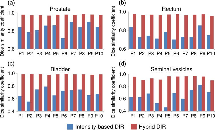
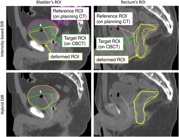
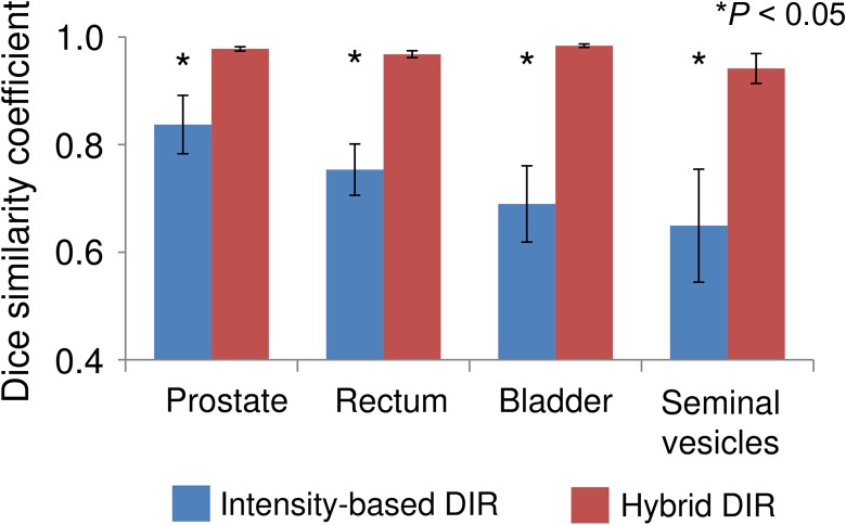
This study aimed to evaluate the performance of the hybrid deformable image registration (DIR) method in comparison with intensity-based DIR for pelvic cone-beam computed tomography (CBCT) images, using intensity and anatomical information. Ten prostate cancer patients treated with intensity-modulated radiation therapy (IMRT) were studied. Nine or ten CBCT scans were performed for each patient. First, rigid registration was performed between the planning CT and all CBCT images using gold fiducial markers, and then DIR was performed. The Dice similarity coefficient (DSC) and center of mass (COM) displacement were used to evaluate the quantitative DIR accuracy. The average DSCs for intensity-based DIR for the prostate, rectum, bladder, and seminal vesicles were 0.84 ± 0.05, 0.75 ± 0.05, 0.69 ± 0.07 and 0.65 ± 0.11, respectively, whereas those values for hybrid DIR were 0.98 ± 0.00, 0.97 ± 0.01, 0.98 ± 0.00 and 0.94 ± 0.03, respectively (P < 0.05). The average COM displacements for intensity-based DIR for the prostate, rectum, bladder, and seminal vesicles were 2.0 ± 1.5, 3.7 ± 1.4, 7.8 ± 2.2 and 3.6 ± 1.2 mm, whereas those values for hybrid DIR were 0.1 ± 0.0, 0.3 ± 0.2, 0.2 ± 0.1 and 0.6 ± 0.6 mm, respectively (P < 0.05). These results showed that the DSC for hybrid DIR had a higher DSC value and smaller COM displacement for all structures and all patients, compared with intensity-based DIR. Thus, the accumulative dose based on hybrid DIR might be trusted as a high-precision dose estimation method that takes into account organ movement during treatment radiotherapy.
本研究旨在评估混合可变形图像配准(DIR)方法与基于强度的DIR方法相比,在使用强度和解剖学信息对盆腔锥形束计算机断层扫描(CBCT)图像进行配准时的性能。对10例接受调强放射治疗(IMRT)的前列腺癌患者进行了研究。每位患者进行了9或10次CBCT扫描。首先,使用金基准标记在计划CT和所有CBCT图像之间进行刚性配准,然后进行DIR。使用骰子相似系数(DSC)和质心(COM)位移来评估定量DIR准确性。基于强度的DIR对前列腺、直肠、膀胱和精囊的平均DSC分别为0.84±0.05、0.75±0.05、0.69±0.07和0.65±0.11,而混合DIR的相应值分别为0.98±0.00、0.97±0.01、0.98±0.00和0.94±0.03(P<0.05)。基于强度的DIR对前列腺、直肠、膀胱和精囊的平均COM位移分别为2.0±1.5、3.7±1.4、7.8±2.2和3.6±1.2mm,而混合DIR的相应值分别为0.1±0.0、0.3±0.2、0.2±0.1和0.6±0.6mm(P<0.05)。这些结果表明,与基于强度的DIR相比,混合DIR的DSC在所有结构和所有患者中具有更高的DSC值和更小的COM位移。因此,基于混合DIR的累积剂量可能被视为一种高精度剂量估计方法,该方法考虑了放射治疗过程中的器官运动。