Neurovirology Laboratory, Department of Medicine, University of Minnesota Medical School, Minneapolis, Minnesota, USA.
Sci Rep. 2017 Feb 6;7:41889. doi: 10.1038/srep41889.
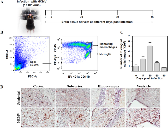
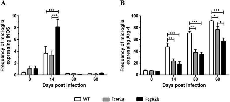
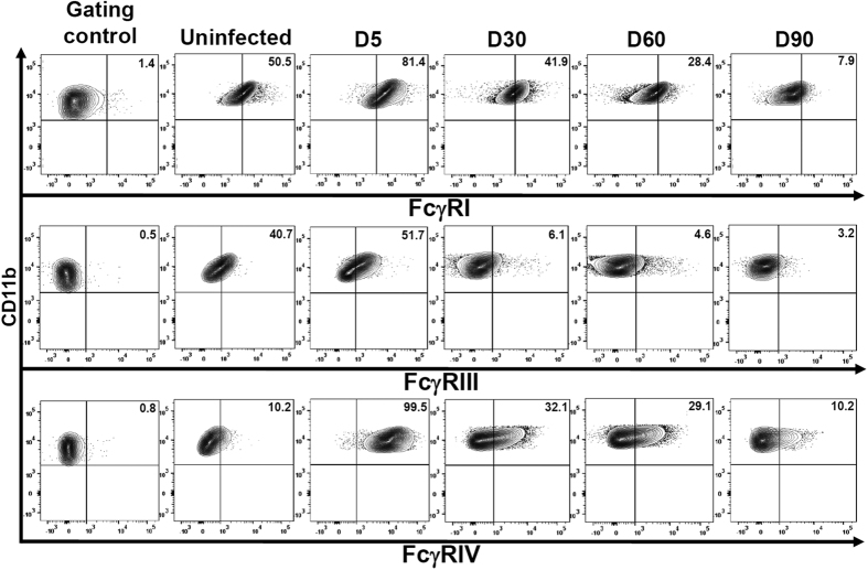
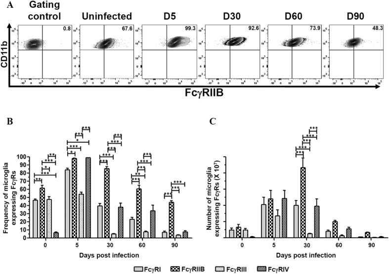
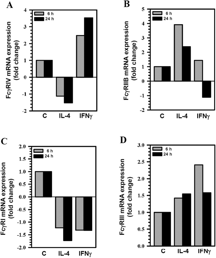
Fcγ receptors (FcγRs) for IgG couple innate and adaptive immunity through activation of effector cells by antigen-antibody complexes. We investigated relative levels of activating and inhibitory FcγRs on brain-resident microglia following murine cytomegalovirus (MCMV) infection. Flow cytometric analysis of microglial cells obtained from infected brain tissue demonstrated that activating FcγRs were expressed maximally at 5 d post-infection (dpi), while the inhibitory receptor (FcγRIIB) remained highly elevated during both acute and chronic phases of infection. The highly induced expression of activating FcγRIV during the acute phase of infection was also noteworthy. Furthermore, in vitro analysis using cultured primary microglia demonstrated the role of interferon (IFN)γ and interleukin (IL)-4 in polarizing these cells towards a M1 or M2 phenotype, respectively. Microglial cell-polarization correlated with maximal expression of either FcγRIV or FcγRIIB following stimulation with IFNγ or IL-4, respectively. Finally, we observed a significant delay in polarization of microglia towards an M2 phenotype in the absence of FcγRs in MCMV-infected Fcer1g and FcgR2b knockout mice. These studies demonstrate that neuro-inflammation following viral infection increases expression of activating FcγRs on M1-polarized microglia. In contrast, expression of the inhibitory FcγRIIB receptor promotes M2-polarization in order to shut-down deleterious immune responses and limit bystander brain damage.
Fcγ 受体(FcγRs)可通过抗原-抗体复合物激活效应细胞,将固有免疫和适应性免疫联系起来。我们研究了鼠巨细胞病毒(MCMV)感染后大脑常驻小胶质细胞上激活和抑制性 FcγRs 的相对水平。对来自感染脑组织的小胶质细胞进行流式细胞术分析表明,激活型 FcγRs 在感染后 5 天(dpi)表达最高,而抑制性受体(FcγRIIB)在感染的急性期和慢性期均高度升高。在感染急性期高度诱导的激活型 FcγRIV 表达也值得注意。此外,使用培养的原代小胶质细胞进行的体外分析表明,干扰素(IFN)γ和白细胞介素(IL)-4 分别在将这些细胞极化为 M1 或 M2 表型方面发挥作用。小胶质细胞极化与 IFNγ或 IL-4 刺激后 FcγRIV 或 FcγRIIB 的最大表达相关。最后,我们观察到在缺乏 FcγRs 的情况下,MCMV 感染的 Fcer1g 和 FcgR2b 基因敲除小鼠中,小胶质细胞向 M2 表型的极化明显延迟。这些研究表明,病毒感染后的神经炎症增加了 M1 极化小胶质细胞上激活型 FcγRs 的表达。相比之下,抑制性 FcγRIIB 受体的表达促进 M2 极化,以关闭有害的免疫反应并限制旁观者脑损伤。