Lee Peter, Ryoo Hojin, Park Jinah, Jeong Yong
Department of Bio and Brain Engineering, Korea Advanced Institute of Science and Technology (KAIST) Daejeon, Korea.
KI for Health Science and Technology, Korea Advanced Institute of Science and Technology (KAIST) Daejeon, Korea.
J Clin Neurol. 2017 Apr;13(2):144-154. doi: 10.3988/jcn.2017.13.2.144. Epub 2017 Jan 25.
With the aim of facilitating the early detection of Alzheimer's disease, the Alzheimer's Disease Neuroimaging Initiative proposed two stages based on the memory performance: early mild cognitive impairment (EMCI) and late mild cognitive impairment (LMCI). The current study was designed to investigate structural differences in terms of surface atrophy and microstructural changes of the hippocampus in EMCI and LMCI.
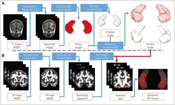
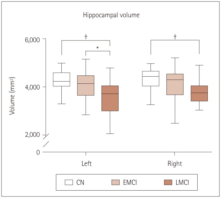
Hippocampal shape modeling based on progressive template surface deformation was performed on T1-weighted MRI images obtained from 20 cognitive normal (CN) subjects, 17 EMCI patients, and 20 LMCI patients. A template surface in CN was used as a region of interest for diffusion-tensor imaging (DTI) voxel-based morphometry (VBM) analysis. Cluster-wise group comparison was performed based on DTI indices within the hippocampus. Linear regression was performed to identify correlations between DTI metrics and clinical scores.
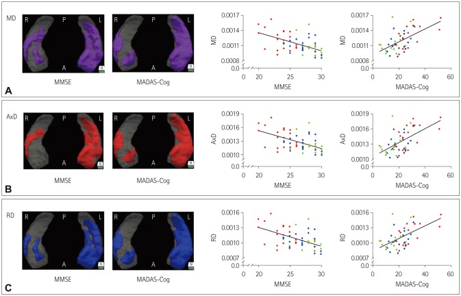
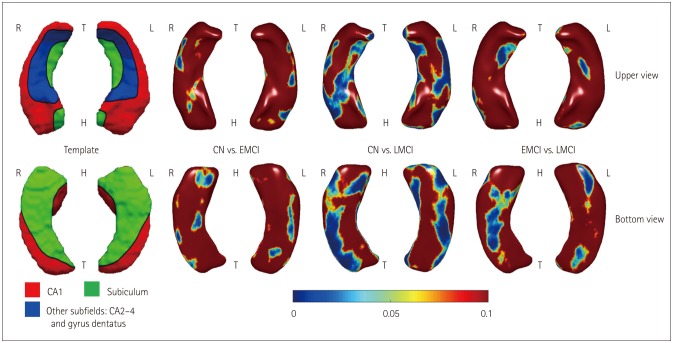
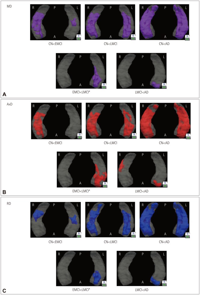
The hippocampal surface analysis showed significant atrophies in bilateral CA1 regions and the right ventral subiculum in EMCI, in contrast to widespread atrophy in LMCI. DTI VBM analysis showed increased diffusivity in the CA2-CA4 regions in EMCI and additionally in the subiculum region in LMCI. Hippocampal diffusivity was significantly correlated with scores both for the Mini Mental State Examination and on the Modified Alzheimer Disease Assessment Scale cognitive subscale. However, the hippocampal diffusivity did not vary significantly with the fractional anisotropy.
EMCI showed hippocampal surface changes mainly in the CA1 region and ventral subiculum. Diffusivity increased mainly in the CA2-CA4 regions in EMCI, while it decreased throughout the hippocampus in LMCI. Although axial diffusivity showed prominent changes in the right hippocampus in EMCI, future studies need to confirm the presence of this laterality difference. In addition, diffusivity is strongly correlated with the cognitive performance, indicating the possibility of using diffusivity as a biomarker for disease progression.
为促进阿尔茨海默病的早期检测,阿尔茨海默病神经影像学倡议组织基于记忆表现提出了两个阶段:早期轻度认知障碍(EMCI)和晚期轻度认知障碍(LMCI)。本研究旨在调查EMCI和LMCI患者海马体表面萎缩和微观结构变化方面的结构差异。
对从20名认知正常(CN)受试者、17名EMCI患者和20名LMCI患者获取的T1加权MRI图像进行基于渐进模板表面变形的海马形状建模。将CN组的模板表面用作基于扩散张量成像(DTI)体素形态计量学(VBM)分析的感兴趣区域。基于海马体内的DTI指标进行聚类组间比较。进行线性回归以确定DTI指标与临床评分之间的相关性。
海马体表面分析显示,EMCI患者双侧CA1区域和右侧腹侧下托有明显萎缩,而LMCI患者则出现广泛萎缩。DTI VBM分析显示,EMCI患者CA2 - CA4区域扩散率增加,LMCI患者另外在海马下托区域扩散率增加。海马体扩散率与简易精神状态检查表评分以及改良阿尔茨海默病评定量表认知子量表评分均显著相关。然而,海马体扩散率与分数各向异性无显著差异。
EMCI主要表现为海马体表面变化,主要在CA1区域和腹侧下托。EMCI患者扩散率主要在CA2 - CA4区域增加,而LMCI患者整个海马体扩散率降低。尽管轴向扩散率在EMCI患者右侧海马体有明显变化,但未来研究需要证实这种左右侧差异的存在。此外,扩散率与认知表现密切相关,表明有可能将扩散率用作疾病进展的生物标志物。