Mkumbaye Sixbert I, Minja Daniel T R, Jespersen Jakob S, Alifrangis Michael, Kavishe Reginald A, Mwakalinga Steven B, Lusingu John P, Theander Thor G, Lavstsen Thomas, Wang Christian W
Kilimanjaro Clinical Research Institute, Kilimanjaro Christian Medical University College, Moshi, Tanzania.
Korogwe Research Station, Tanga Centre, National Institute for Medical Research, Tanga, Tanzania.
Malar J. 2017 Feb 10;16(1):69. doi: 10.1186/s12936-017-1714-2.
Establishing in vitro Plasmodium falciparum culture lines from patient parasite isolates can offer deeper understanding of geographic variations of drug sensitivity and mechanisms of malaria pathogenesis and immunity. Cellulose column filtration of blood is an inexpensive, rapid and effective method for the removal of host factors, such as leucocytes and platelets, significantly improving the purification of parasite DNA in a blood sample.
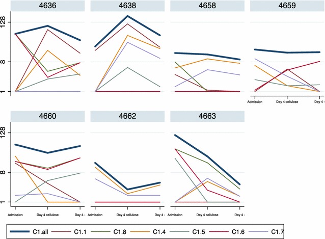
In this study, the effect of cellulose column filtration of venous blood on the initial in vitro growth of P. falciparum parasite isolates from Tanzanian children admitted to hospital was tested. The parasites were allowed to expand in culture without subcultivation until 5 days after admission or the appearance of dead parasites and parasitaemia was determined daily. To investigate whether the filtration had an effect on clonality, P. falciparum merozoite surface protein 2 genotyping was performed using nested PCR on extracted genomic DNA, and the var gene transcript levels were investigated, using quantitative PCR on extracted RNA, at admission and 4 days of culture.
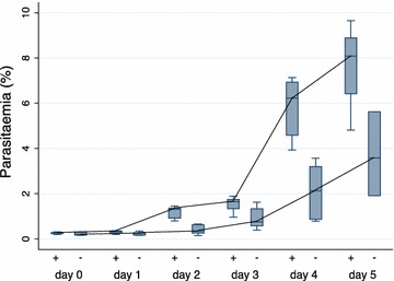
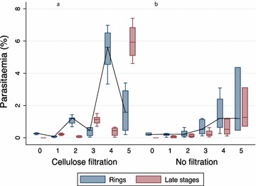
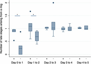
The cellulose-filtered parasites grew to higher parasitaemia faster than non-filtered parasites seemingly due to a higher development ratio of ring stage parasites progressing into the late stages. Cellulose filtration had no apparent effect on clonality or var gene expression; however, evident differences were observed after only 4 days of culture in both the number of clones and transcript levels of var genes compared to the time of admission.
Cellulose column filtration of parasitized blood is a cheap, applicable method for improving cultivation of P. falciparum field isolates for ex vivo based assays; however, when assessing phenotype and genotype of cultured parasites, in general, assumed to represent the in vivo infection, caution is advised.
从患者寄生虫分离株建立恶性疟原虫体外培养系,有助于更深入地了解药物敏感性的地理差异以及疟疾发病机制和免疫机制。血液的纤维素柱过滤是一种廉价、快速且有效的方法,可去除宿主因子,如白细胞和血小板,显著提高血样中寄生虫DNA的纯度。
在本研究中,测试了静脉血的纤维素柱过滤对坦桑尼亚住院儿童恶性疟原虫分离株体外初始生长的影响。让寄生虫在培养中扩增而不进行传代培养,直到入院后5天或出现死亡寄生虫,并每天测定疟原虫血症。为了研究过滤是否对克隆性有影响,对提取的基因组DNA进行巢式PCR,以进行恶性疟原虫裂殖子表面蛋白2基因分型,并在入院时和培养4天时,对提取的RNA进行定量PCR,以研究var基因转录水平。
纤维素过滤的寄生虫比未过滤的寄生虫更快地生长到更高的疟原虫血症水平,这似乎是由于环状期寄生虫向后期发展的比例更高。纤维素过滤对克隆性或var基因表达没有明显影响;然而,与入院时相比,仅在培养4天后,在克隆数量和var基因转录水平上就观察到了明显差异。
寄生血液的纤维素柱过滤是一种廉价、适用的方法,可改善用于体外检测的恶性疟原虫野外分离株的培养;然而,在评估培养寄生虫的表型和基因型时,通常假定其代表体内感染,建议谨慎行事。