Sjöberg Pia, Heiberg Einar, Wingren Pär, Ramgren Johansson Jens, Malm Torsten, Arheden Håkan, Liuba Petru, Carlsson Marcus
Department of Clinical Sciences Lund, Clinical Physiology, Skane University Hospital, Lund University, Lund, Sweden.
Department of Medical Imaging and Physiology, Skane University Hospital, Lund, Sweden.
Pediatr Cardiol. 2017 Apr;38(4):669-680. doi: 10.1007/s00246-016-1565-6. Epub 2017 Feb 10.
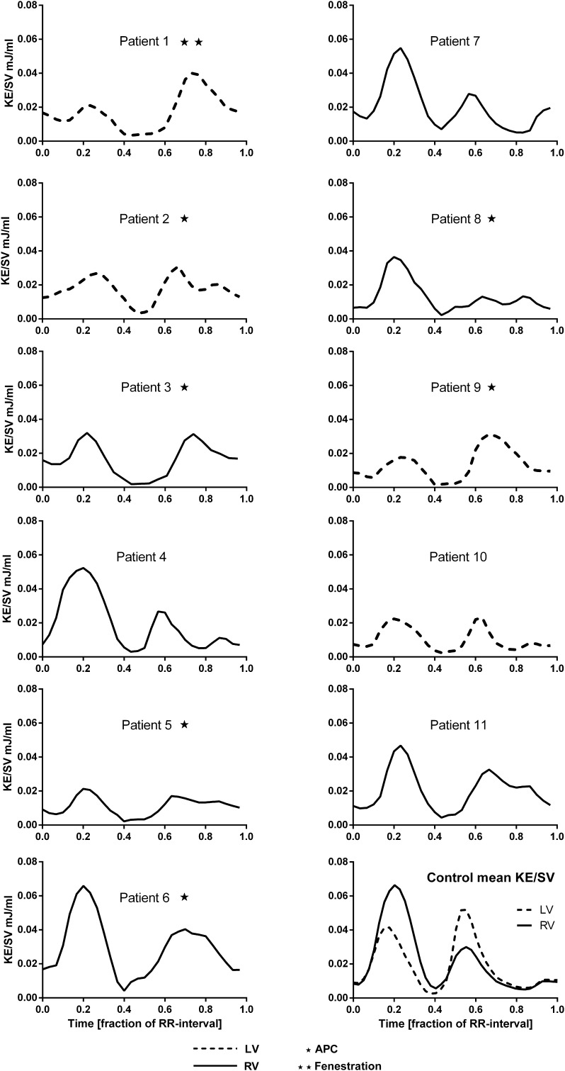
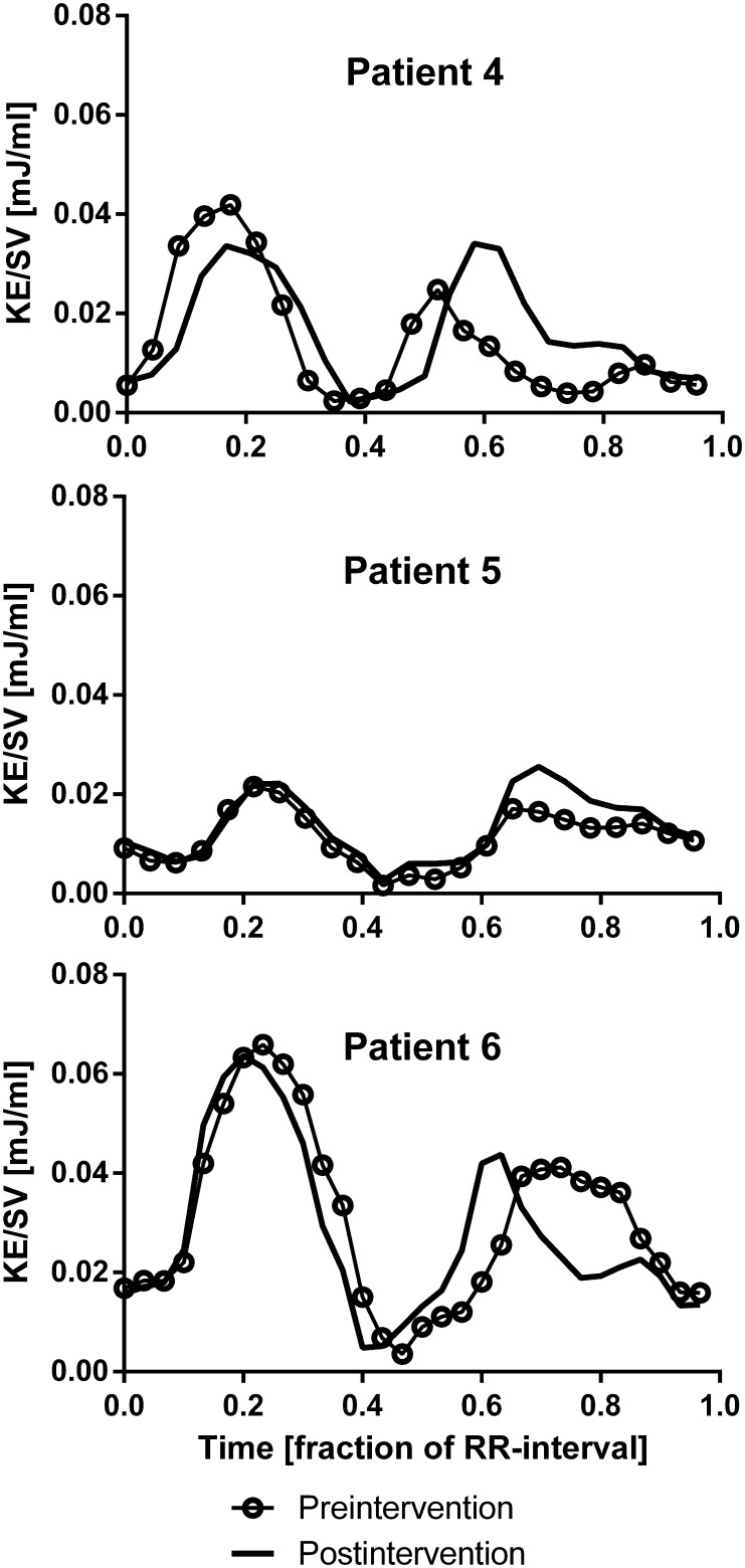
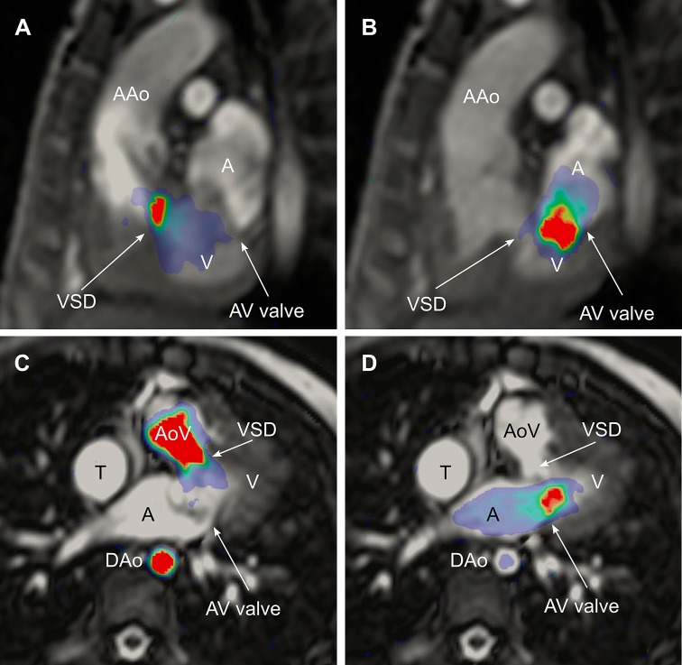
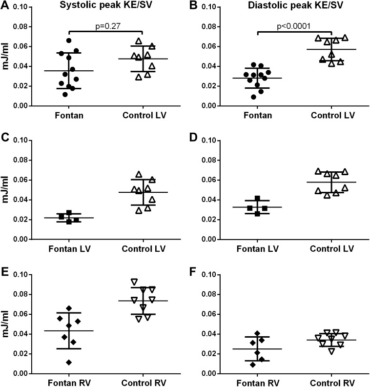
Four-dimensional (4D) flow magnetic resonance imaging (MRI) enables quantification of kinetic energy (KE) in intraventricular blood flow. This provides a novel way to understand the cardiovascular physiology of the Fontan circulation. In this study, we aimed to quantify the KE in functional single ventricles. 4D flow MRI was acquired in eleven patients with Fontan circulation (median age 12 years, range 3-29) and eight healthy volunteers (median age 26 years, range 23-36). Follow-up MRI after surgical or percutaneous intervention was performed in 3 patients. Intraventricular KE was calculated throughout the cardiac cycle and indexed to stroke volume (SV). The systolic/diastolic ratio of KE in Fontan patients was similar to the ratio of the controls' left ventricle (LV) or right ventricle (RV) depending on the patients' ventricular morphology (Cohen´s κ = 1.0). Peak systolic KE/SV did not differ in patients compared to the LV in controls (0.016 ± 0.006 mJ/ml vs 0.020 ± 0.004 mJ/ml, p = 0.09). Peak diastolic KE/SV in Fontan patients was lower than in the LV of the control group (0.028 ± 0.010 mJ/ml vs 0.057 ± 0.011 mJ/ml, p < 0.0001). The KE during diastole showed a plateau in patients with aortopulmonary collaterals. This is to our knowledge the first study that quantifies the intraventricular KE of Fontan patients. KE is dependent on the morphology of the ventricle, and diastolic KE indexed to SV in patients is decreased compared to controls. The lower KE in Fontan patients may be a result of impaired ventricular filling.
四维(4D)血流磁共振成像(MRI)能够对脑室内血流的动能(KE)进行量化。这为理解Fontan循环的心血管生理学提供了一种新方法。在本研究中,我们旨在量化功能性单心室中的KE。对11例Fontan循环患者(中位年龄12岁,范围3 - 29岁)和8名健康志愿者(中位年龄26岁,范围23 - 36岁)进行了4D血流MRI检查。3例患者在手术或经皮介入后进行了随访MRI检查。在整个心动周期中计算脑室内KE,并将其与每搏量(SV)进行指数化。Fontan患者KE的收缩期/舒张期比值与对照组左心室(LV)或右心室(RV)的比值相似,具体取决于患者的心室形态(Cohen's κ = 1.0)。与对照组LV相比,患者的收缩期峰值KE/SV无差异(0.016±0.006 mJ/ml对0.020±0.004 mJ/ml,p = 0.09)。Fontan患者的舒张期峰值KE/SV低于对照组LV(0.028±0.010 mJ/ml对0.057±0.011 mJ/ml,p < 0.0001)。有主肺动脉侧支的患者在舒张期KE呈现平台期。据我们所知,这是第一项量化Fontan患者脑室内KE的研究。KE取决于心室形态,与对照组相比,患者中以SV指数化的舒张期KE降低。Fontan患者较低的KE可能是心室充盈受损的结果。