Wilson J M, Mukherjee S, Brunner T B, Partridge M, Hawkins M A
CRUK/MRC Oxford Institute for Radiation Oncology, Gray Laboratories, Oxford, UK.
CRUK/MRC Oxford Institute for Radiation Oncology, Gray Laboratories, Oxford, UK.
Clin Oncol (R Coll Radiol). 2017 Jun;29(6):370-377. doi: 10.1016/j.clon.2017.01.038. Epub 2017 Feb 9.
A proportion of patients with pancreatic cancer never develop metastatic disease. We evaluated a role for F-fluorodeoxyglucose positron emission tomography (FDG-PET) in identifying a subset of patients with locally advanced pancreatic cancer (LAPC) who never develop metastatic disease and only experience local disease and may therefore benefit from local treatment intensification.
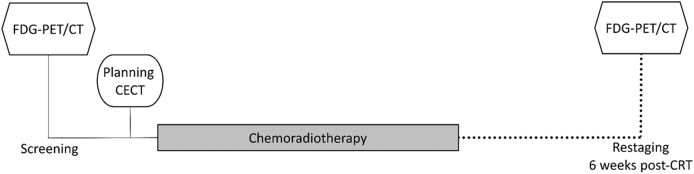
Patients with histologically confirmed LAPC entered a single-centre phase II study of definitive upfront chemoradiotherapy (CRT). All patients underwent FDG-PET/CT before and 6 weeks after CRT. Tumour volume, standardised uptake values (SUV, SUV, SUV, SUV) and total lesion glycolysis (TLG) were measured on each scan and the response in each parameter was evaluated. The presence or absence of metastatic disease was noted on contrast-enhanced CT carried out every 3 months for 1 year and then at clinician discretion.
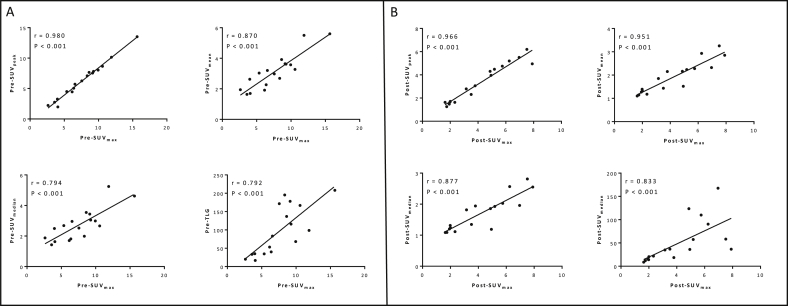
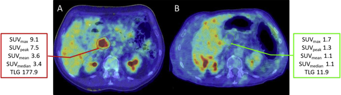
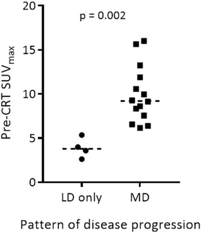
Twenty-three patients with LAPC were recruited; 17/23 completed treatment and had interpretable sequential imaging. Twenty-four per cent of patients only ever experienced local disease. Median pre-CRT FDG-PET parameters were significantly lower in patients with local disease only during follow-up compared with those who developed metastatic disease: SUV 3.8 versus 8.6 (P=0.006), SUV 2.5 versus 7.5 (P=0.002), SUV 1.8 versus 3.3 (P=0.001), SUV 1.7 versus 3.0 (P=0.002), TLG 26.9 versus 115.9 (P=0.006). Tumour volume, post-CRT FDG-PET values and their relative change were not statistically different between local disease and metastatic disease groups. Receiver operating characteristic curves for pre-CRT FDG-PET parameters to predict those who never develop metastatic disease all had areas under the curve (AUCs) ≥ 0.932. Pre-CRT FDG-PET SUV < 6.2 predicted patients with local disease only during follow-up with 100.0% sensitivity and 92.3% specificity, 80.0% positive predictive value and 100% negative predictive value.
Our findings suggest that patients with less FDG-avid tumours are less likely to metastasise and may therefore benefit from upfront local treatment intensification.
一部分胰腺癌患者从未发生转移。我们评估了F-氟脱氧葡萄糖正电子发射断层扫描(FDG-PET)在识别局部晚期胰腺癌(LAPC)患者亚组中的作用,这些患者从未发生转移,仅出现局部病变,因此可能从强化局部治疗中获益。
组织学确诊为LAPC的患者进入一项确定性 upfront 放化疗(CRT)的单中心 II 期研究。所有患者在CRT前和CRT后6周均接受FDG-PET/CT检查。在每次扫描时测量肿瘤体积、标准化摄取值(SUV、SUV、SUV、SUV)和总病变糖酵解(TLG),并评估每个参数的反应。在1年中每3个月进行一次对比增强CT检查,之后由临床医生酌情决定,记录是否存在转移。
招募了23例LAPC患者;17/23例完成治疗并具有可解释的序贯成像。24%的患者仅出现局部病变。仅出现局部病变的患者在随访期间,CRT前FDG-PET参数的中位数显著低于发生转移的患者:SUV 3.8对8.6(P = 0.006),SUV 2.5对7.5(P = 0.002),SUV 1.8对3.3(P = 0.001),SUV 1.7对3.0(P = 0.002),TLG 26.9对115.9(P = 0.006)。局部病变组和转移组之间,肿瘤体积、CRT后FDG-PET值及其相对变化无统计学差异。用于预测从未发生转移患者的CRT前FDG-PET参数的受试者工作特征曲线,曲线下面积(AUC)均≥0.932。CRT前FDG-PET SUV < 6.2预测仅在随访期间出现局部病变的患者,敏感性为100.0%,特异性为92.3%,阳性预测值为80.0%,阴性预测值为100%。
我们的研究结果表明,FDG摄取较低的肿瘤患者发生转移的可能性较小,因此可能从 upfront 局部治疗强化中获益。