Benharroch Daniel, Zekzer Miriam, Nalbandyan Karen
Department of Pathology, Soroka University Medical Center and Faculty of Health Sciences, Ben-Gurion University of the Negev, Beer-Sheva, Israel.
Department of Hematology, Soroka University Medical Center and Faculty of Health Sciences, Ben-Gurion University of the Negev, Beer-Sheva, Israel.
Case Rep Hematol. 2017;2017:9601094. doi: 10.1155/2017/9601094. Epub 2017 Jan 18.
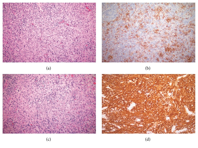
An elderly woman presented with generalized lymphadenopathy, several systemic symptoms, and splenomegaly. An inguinal lymph node excision revealed a compound picture. One aspect of the lymph node morphology, including cells with follicular T-helper cell phenotype, was most consistent with angioimmunoblastic T-cell lymphoma. The other component, revealing spindle cells forming whorls with immunostaining for CD21, CD23, and fascin, might be an integral part of this T-cell lymphoma. However, due to the often massive involvement of the nodal tissue by these follicular dendritic cells, these areas were questionably suggestive of involvement by follicular dendritic cell sarcoma. We raise herein the issue of the borderline area between advanced follicular dendritic cell expansion in angioimmunoblastic T-cell lymphoma and a massive follicular dendritic cell proliferation consistent with follicular dendritic cells sarcoma, in the absence of a genomic analysis.
一名老年女性出现全身淋巴结肿大、多种全身症状及脾肿大。腹股沟淋巴结切除显示出复合表现。淋巴结形态的一个方面,包括具有滤泡性辅助性T细胞表型的细胞,最符合血管免疫母细胞性T细胞淋巴瘤。另一个成分显示梭形细胞形成漩涡状,CD21、CD23和fascin免疫染色阳性,可能是这种T细胞淋巴瘤的一个组成部分。然而,由于这些滤泡树突状细胞常大量累及淋巴结组织,这些区域可疑提示滤泡树突状细胞肉瘤累及。在缺乏基因组分析的情况下,我们在此提出血管免疫母细胞性T细胞淋巴瘤中晚期滤泡树突状细胞扩张与符合滤泡树突状细胞肉瘤的大量滤泡树突状细胞增殖之间的临界区域问题。