McNally Andrew, Madan Ashish, Sucosky Philippe
Department of Aerospace and Mechanical Engineering, University of Notre Dame Notre Dame, IN, USA.
Department of Mechanical and Materials Engineering, Wright State University Dayton, OH, USA.
Front Physiol. 2017 Feb 1;8:44. doi: 10.3389/fphys.2017.00044. eCollection 2017.
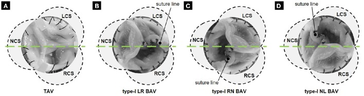
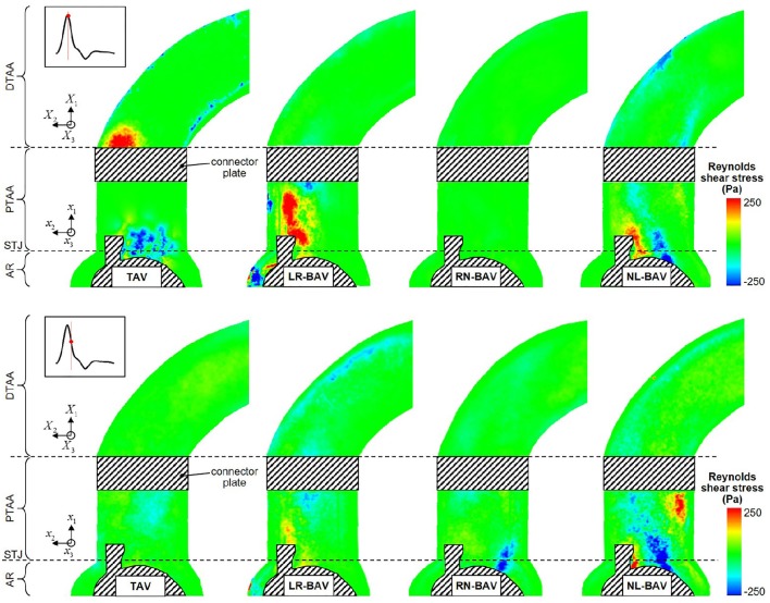
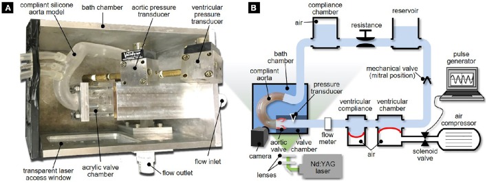
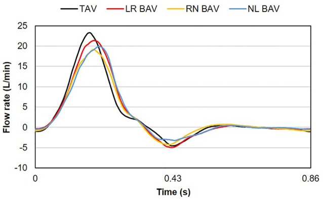
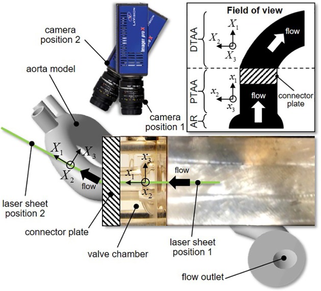
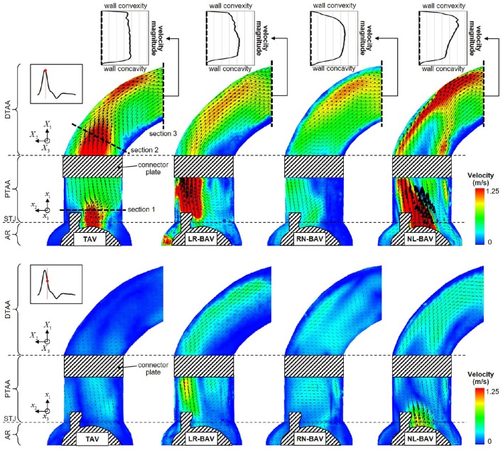
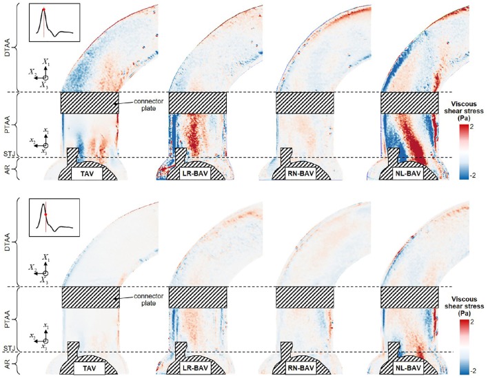
The bicuspid aortic valve (BAV) is a major risk factor for secondary aortopathy such as aortic dilation. The heterogeneous BAV morphotypes [left-right-coronary cusp fusion (LR), right-non-coronary cusp fusion (RN), and left-non-coronary cusp fusion (LN)] are associated with different dilation patterns, suggesting a role for hemodynamics in BAV aortopathogenesis. However, assessment of this theory is still hampered by the limited knowledge of the hemodynamic abnormalities generated by the distinct BAV morphotypes. The objective of this study was to compare experimentally the hemodynamics of a normal (i.e., non-dilated) ascending aorta (AA) subjected to tricuspid aortic valve (TAV), LR-BAV, RN-BAV, and NL-BAV flow. Tissue BAVs reconstructed from porcine TAVs were subjected to physiologic pulsatile flow conditions in a left-heart simulator featuring a realistic aortic root and compliant aorta. Phase-locked particle image velocimetry experiments were carried out to characterize the flow in the aortic root and in the tubular AA in terms of jet skewness and displacement, as well as mean velocity, viscous shear stress and Reynolds shear stress fields. While all three BAVs generated skewed and asymmetrical orifice jets (up to 1.7- and 4.0-fold increase in flow angle and displacement, respectively, relative to the TAV at the sinotubular junction), the RN-BAV jet was out of the plane of observation. The LR- and NL-BAV exhibited a 71% increase in peak-systolic orifice jet velocity relative to the TAV, suggesting an inherent degree of stenosis in BAVs. While these two BAV morphotypes subjected the convexity of the aortic wall to viscous shear stress overloads (1.7-fold increase in maximum peak-systolic viscous shear stress relative to the TAV-AA), the affected sites were morphotype-dependent (LR-BAV: proximal AA, NL-BAV: distal AA). Lastly, the LR- and NL-BAV generated high degrees of turbulence in the AA (up to 2.3-fold increase in peak-systolic Reynolds shear stress relative to the TAV) that were sustained from peak systole throughout the deceleration phase. This study reveals substantial flow abnormalities (increased jet skewness, asymmetry, jet velocity, turbulence, and shear stress overloads) in non-dilated BAV aortas, which differ from those observed in dilated aortas but still coincide with aortic wall regions prone to dilation.
二叶式主动脉瓣(BAV)是继发性主动脉病变(如主动脉扩张)的主要危险因素。不同类型的BAV形态(左右冠状动脉瓣叶融合型(LR)、右无冠状动脉瓣叶融合型(RN)和左无冠状动脉瓣叶融合型(LN))与不同的扩张模式相关,提示血流动力学在BAV主动脉病变发生过程中起作用。然而,由于对不同BAV形态所产生的血流动力学异常了解有限,该理论的评估仍受到阻碍。本研究的目的是通过实验比较正常(即未扩张)升主动脉(AA)在三尖瓣主动脉瓣(TAV)、LR-BAV、RN-BAV和NL-BAV血流作用下的血流动力学。从猪TAV重建的组织BAV在具有逼真主动脉根部和顺应性主动脉的左心模拟器中接受生理脉动血流条件。进行锁相粒子图像测速实验,以根据射流偏斜度和位移以及平均速度、粘性剪切应力和雷诺剪切应力场来表征主动脉根部和管状AA中的血流。虽然所有三种BAV均产生偏斜且不对称的瓣口射流(相对于窦管交界处的TAV,血流角度和位移分别增加高达1.7倍和4.0倍),但RN-BAV射流不在观察平面内。相对于TAV,LR-BAV和NL-BAV的收缩期峰值瓣口射流速度增加了71%,提示BAV存在固有程度的狭窄。虽然这两种BAV形态使主动脉壁凸面承受粘性剪切应力过载(相对于TAV-AA,最大收缩期峰值粘性剪切应力增加1.7倍),但受影响部位取决于形态类型(LR-BAV:升主动脉近端,NL-BAV:升主动脉远端)。最后,LR-BAV和NL-BAV在AA中产生高度湍流(相对于TAV,收缩期峰值雷诺剪切应力增加高达2.3倍),从收缩期峰值持续到减速期。本研究揭示了未扩张的BAV主动脉中存在大量血流异常(射流偏斜度增加、不对称、射流速度、湍流和剪切应力过载),这些异常与扩张主动脉中观察到的不同,但仍与易于扩张的主动脉壁区域一致。