Piatti Filippo, Sturla Francesco, Bissell Malenka M, Pirola Selene, Lombardi Massimo, Nesteruk Igor, Della Corte Alessandro, Redaelli Alberto C L, Votta Emiliano
Department of Electronics, Information and Bioengineering, Politecnico di MilanoMilan, Italy.
Division of Cardiovascular Medicine, Radcliffe Department of Medicine, University of OxfordOxford, United Kingdom.
Front Physiol. 2017 Jun 26;8:441. doi: 10.3389/fphys.2017.00441. eCollection 2017.
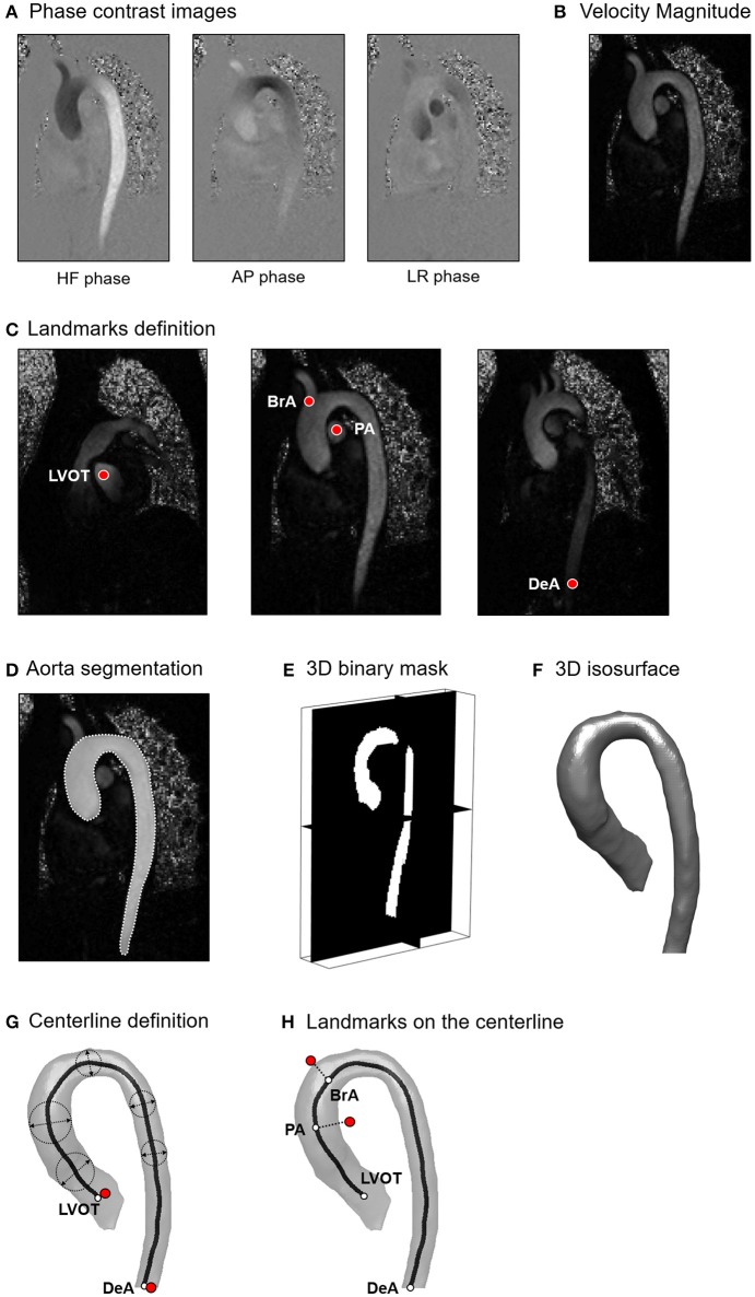
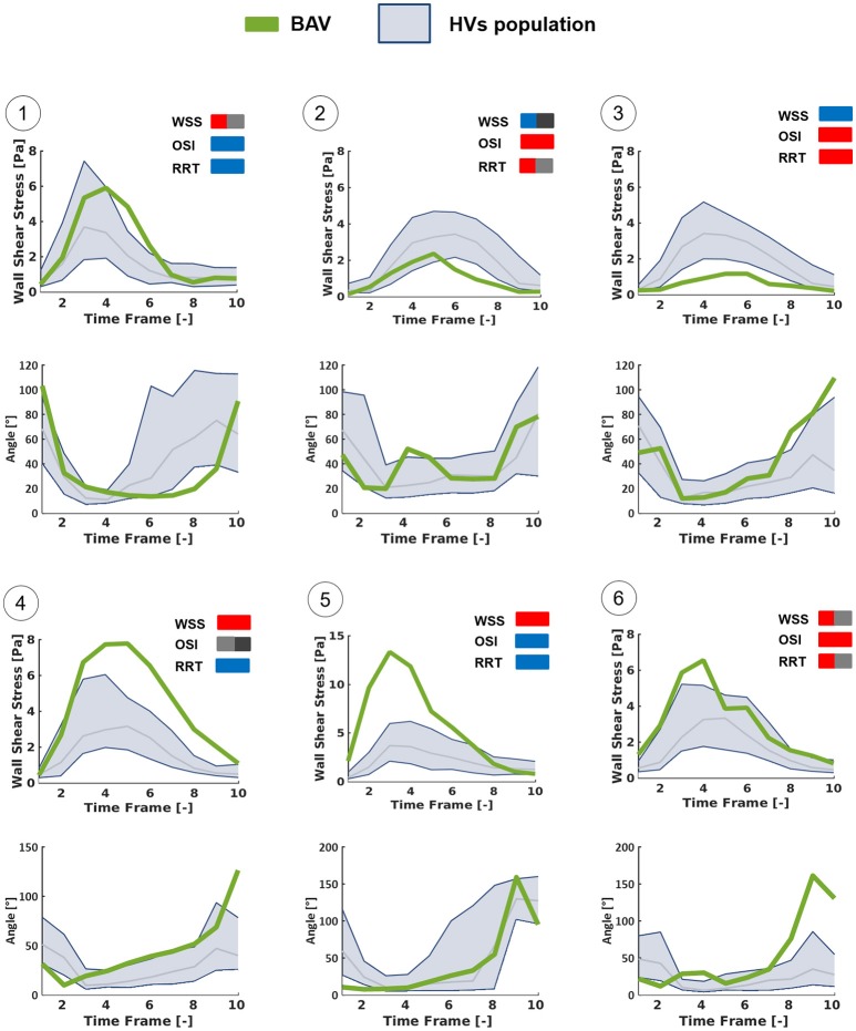
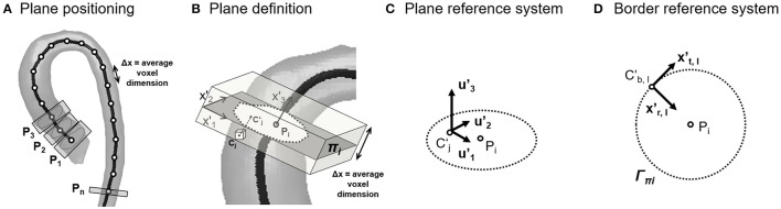
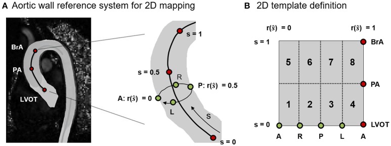
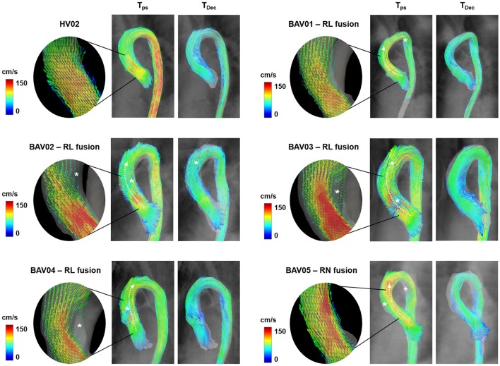
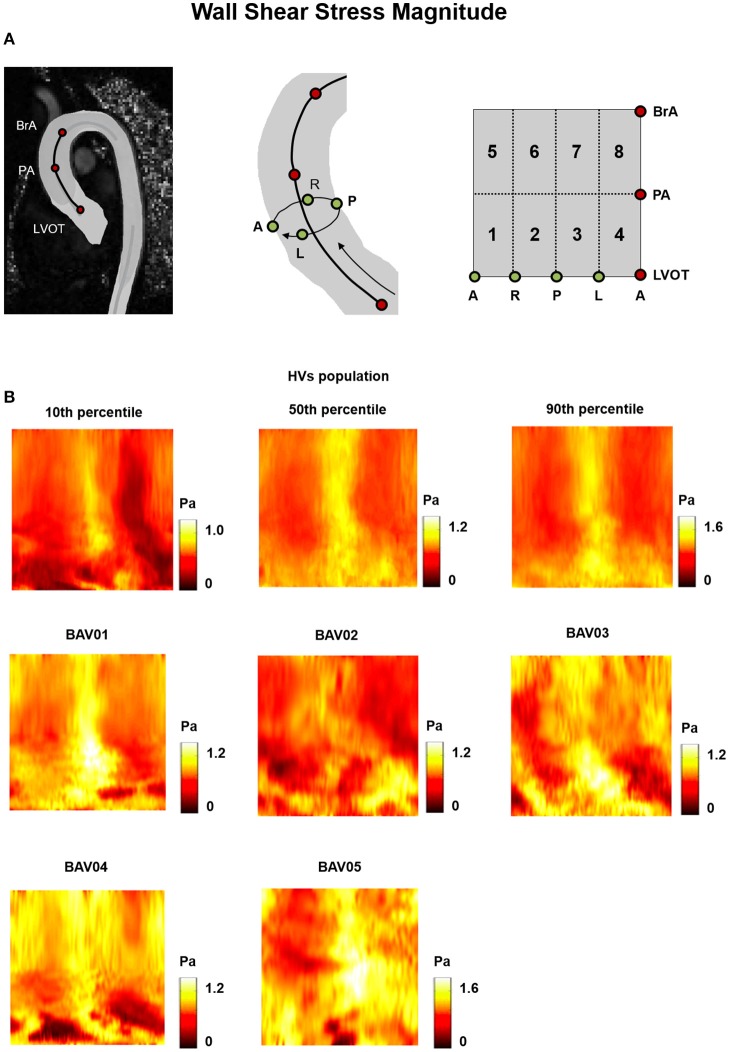
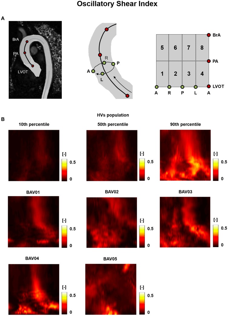
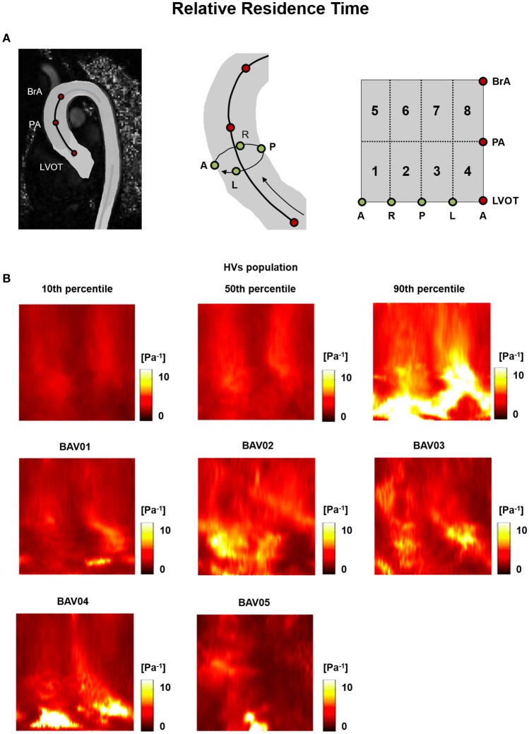
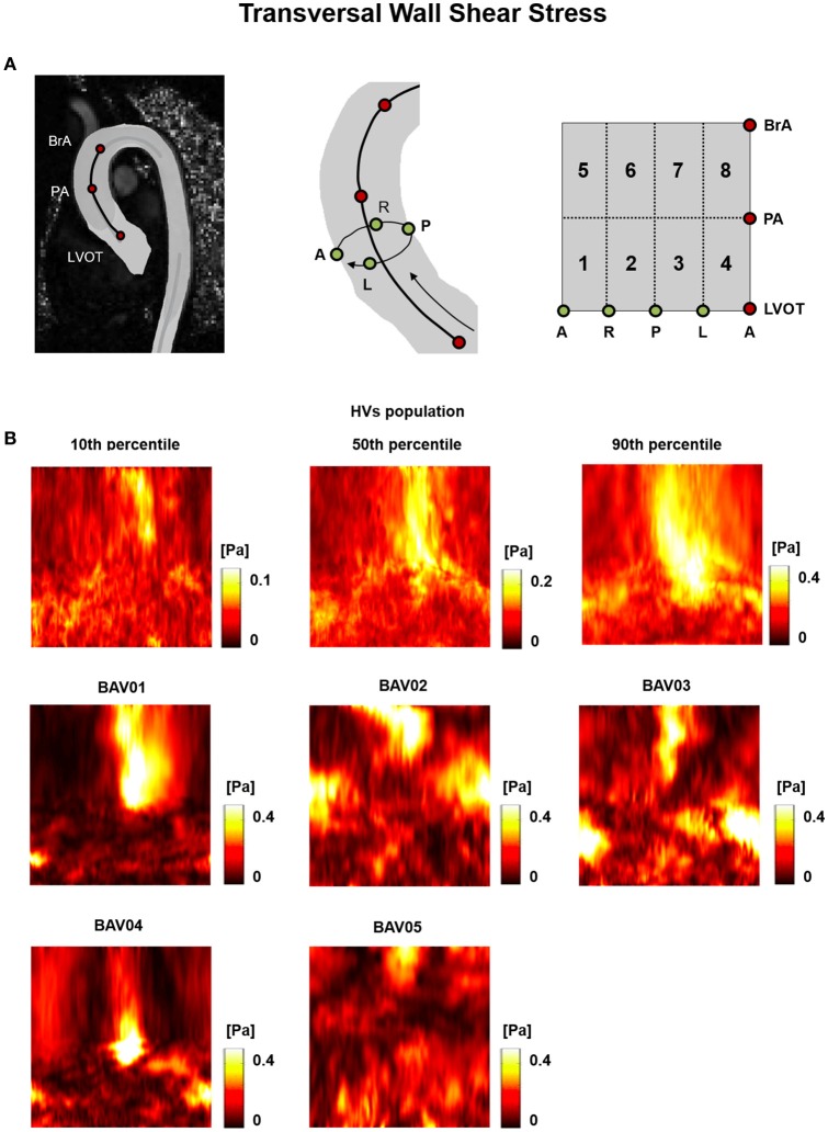
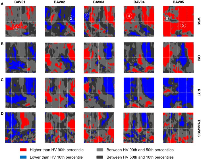
Bicuspid aortic valve (BAV) is the most common congenital cardiac disease and is a foremost risk factor for aortopathies. Despite the genetic basis of BAV and of the associated aortopathies, BAV-related alterations in aortic fluid-dynamics, and particularly in wall shear stresses (WSSs), likely play a role in the progression of aortopathy, and may contribute to its pathogenesis. To test whether WSS may trigger aortopathy, in this study we used 4D Flow sequences of phase-contrast cardiac magnetic resonance imaging (CMR) to quantitatively compare the fluid dynamics in the thoracic aorta of two groups of subjects: (i) five prospectively enrolled young patients with normo-functional BAV and with no aortic dilation and (ii) ten age-matched healthy volunteers. Through the semi-automated processing of 4D Flow data, the aortic bulk flow at peak systole was quantified, and WSSs acting on the endothelium of the ascending aorta were characterized throughout the systolic phase in terms of magnitude and time-dependency through a method recently developed by our group. Variables computed for each BAV patient were compared vs. the corresponding distribution of values obtained for healthy controls. In BAV patients, ascending aorta diameter was measured on cine-CMR images at baseline and at 3-year follow-up. As compared to controls, normo-functional BAV patients were characterized by minor bulk flow disturbances at peak systole. However, they were characterized by evident alterations of WSS distribution and peak values in the ascending aorta. In particular, in four BAV patients, who were characterized by right-left leaflet fusion, WSS peak values exceeded by 27-46% the 90th percentile of the distribution obtained for healthy volunteers. Only in the BAV patient with right-non-coronary leaflet fusion the same threshold was exceeded by 132%. Also, evident alterations in the time-dependency of WSS magnitude and direction were observed. Despite, these fluid-dynamic alterations, no clinically relevant anatomical remodeling was observed in the BAV patients at 3-year follow-up. In light of previous evidence from the literature, our results suggest that WSS alterations may precede the onset of aortopathy and may contribute to its triggering, but WSS-driven anatomical remodeling, if any, is a very slow process.
二叶式主动脉瓣(BAV)是最常见的先天性心脏病,也是主动脉病变的首要危险因素。尽管BAV及相关主动脉病变存在遗传基础,但BAV相关的主动脉流体动力学改变,尤其是壁面剪应力(WSSs),可能在主动脉病变进展中起作用,并可能促成其发病机制。为了测试WSS是否会引发主动脉病变,在本研究中,我们使用相位对比心脏磁共振成像(CMR)的4D流序列,定量比较两组受试者胸主动脉的流体动力学:(i)五名前瞻性招募的具有正常功能BAV且无主动脉扩张的年轻患者,以及(ii)十名年龄匹配的健康志愿者。通过对4D流数据的半自动处理,量化了收缩末期的主动脉总体血流量,并通过我们小组最近开发的一种方法,在整个收缩期对作用于升主动脉内皮的WSSs的大小和时间依赖性进行了表征。将为每位BAV患者计算的变量与健康对照者获得的相应值分布进行比较。在BAV患者中,在基线和3年随访时通过电影CMR图像测量升主动脉直径。与对照组相比,具有正常功能的BAV患者在收缩末期的总体血流紊乱较小。然而,他们的特征是升主动脉中WSS分布和峰值明显改变。特别是,在四名以左右瓣叶融合为特征的BAV患者中,WSS峰值超过健康志愿者分布的第90百分位数27%-46%。只有在以右非冠状动脉瓣叶融合为特征的BAV患者中,同一阈值超过了132%。此外,还观察到WSS大小和方向的时间依赖性明显改变。尽管存在这些流体动力学改变,但在3年随访中,BAV患者未观察到临床相关的解剖重塑。根据先前文献中的证据,我们的结果表明,WSS改变可能先于主动脉病变的发生,并可能促成其触发,但WSS驱动的解剖重塑(如果有的话)是一个非常缓慢的过程。