Li Yun, Mei Zhu, Liu Shuiqiao, Wang Tong, Li Hui, Li Xiao-Xiao, Han Song, Yang Yutao, Li Junfa, Xu Zhi-Qing David
Department of Neurobiology and Beijing Institute for Brain Disorders, Capital Medical University, Beijing 100069, China.
Aging Dis. 2017 Feb 1;8(1):85-100. doi: 10.14336/AD.2016.0806. eCollection 2017 Feb.
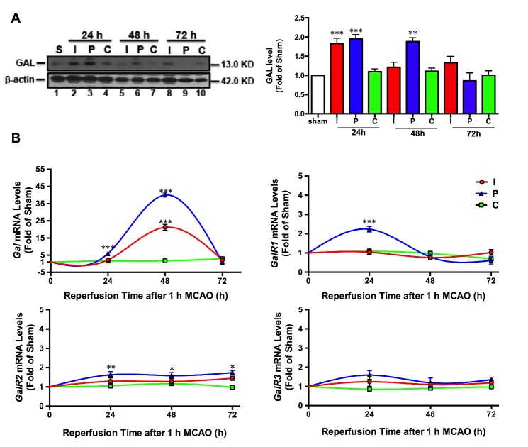
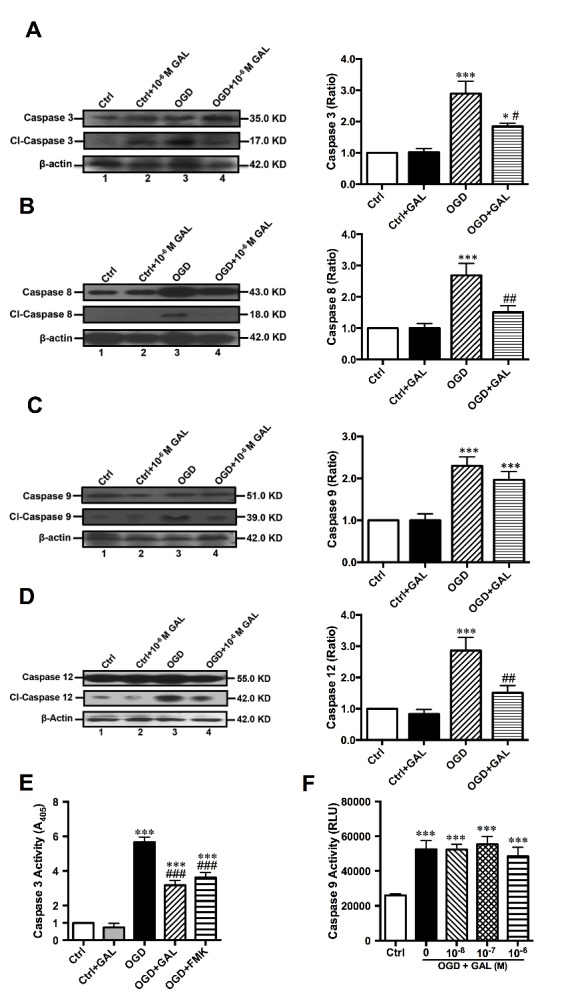
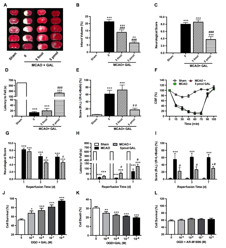
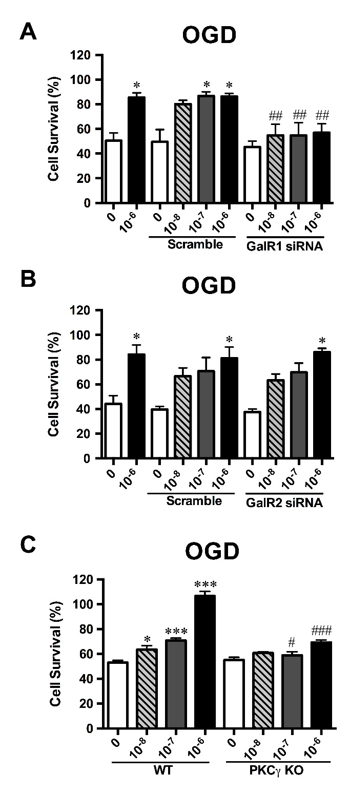
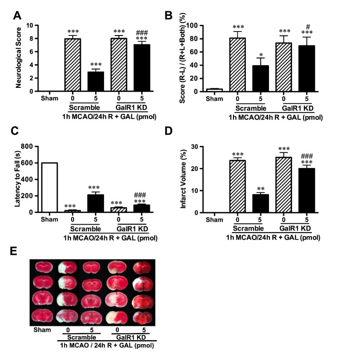
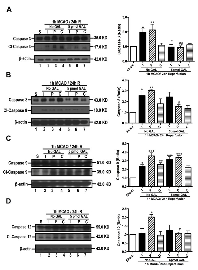
Galanin (GAL) plays key role in many pathophysiological processes, but its role in ischemic stroke remains unclear. Here, the models of 1 h middle cerebral artery occlusion (MCAO)/1-7 d reperfusion (R)-induced ischemic stroke and cell ischemia of 1 h oxygen-glucose deprivation (OGD)/24 h reoxygenation in primary cultured cortical neurons were used to explore GAL's effects and its underlying mechanisms. The results showed significant increases of GAL protein levels in the peri-infarct region (P) and infarct core (I) within 48 h R of MCAO mice (<0.001). The RT-qPCR results also demonstrated significant increases of mRNA during 24-48 h R (<0.001), and GAL receptors (but not 3) mRNA levels in the P region at 24 h R of MCAO mice (<0.001). Furthermore, the significant decrease of infarct volume (<0.05) and improved neurological outcome (<0.001-0.05) were observed in MCAO mice following 1 h pre- or 6 h post-treatment of GAL during 1-7 d reperfusion. GalR1 was confirmed as the receptor responsible for GAL-induced neuroprotection by using GalR2/3 agonist AR-M1896 and Lentivirus-based RNAi knockdown of GalR1. GAL treatment inhibited Caspase-3 activation through the upstream initiators Capsases-8/-12 (not Caspase-9) in both P region and OGD-treated cortical neurons. Meanwhile, GAL's neuroprotective effect was not observed in cortical neurons from conventional protein kinase C (cPKC) γ knockout mice. These results suggested that exogenous GAL protects the brain from ischemic injury by inhibiting Capsase-8/12-initiated apoptosis, possibly mediated by GalR1 via the cPKCγ signaling pathway.
甘丙肽(GAL)在许多病理生理过程中起关键作用,但其在缺血性脑卒中中的作用仍不清楚。在此,采用1小时大脑中动脉闭塞(MCAO)/1 - 7天再灌注(R)诱导的缺血性脑卒中模型以及原代培养皮质神经元1小时氧糖剥夺(OGD)/24小时复氧的细胞缺血模型,以探究GAL的作用及其潜在机制。结果显示,在MCAO小鼠再灌注48小时内,梗死灶周围区域(P)和梗死核心(I)的GAL蛋白水平显著升高(<0.001)。RT-qPCR结果还表明,在再灌注24 - 48小时期间mRNA显著增加(<0.001),且在MCAO小鼠再灌注24小时时,P区域的GAL受体(但不包括3)mRNA水平显著增加(<0.001)。此外,在再灌注1 - 7天期间,对MCAO小鼠进行GAL预处理1小时或后处理6小时后,观察到梗死体积显著减小(<0.05),神经功能结局得到改善(<0.001 - 0.05)。通过使用GalR2/3激动剂AR-M1896和基于慢病毒的GalR1 RNAi敲低,证实GalR1是负责GAL诱导神经保护作用的受体。GAL处理在P区域和OGD处理的皮质神经元中均通过上游起始因子Caspases-8/-12(而非Caspase-9)抑制Caspase-3的激活。同时,在传统蛋白激酶C(cPKC)γ基因敲除小鼠的皮质神经元中未观察到GAL的神经保护作用。这些结果表明,外源性GAL通过抑制Caspase-8/12启动的细胞凋亡保护大脑免受缺血性损伤,可能是由GalR1经由cPKCγ信号通路介导的。