Obata Yurie, Ong Qi J, Magruder J T, Grichkevitch Helen, Berkowitz Dan E, Nyhan Daniel, Steppan Jochen, Barodka Viachaslau
Division of Cardiac Anesthesia, Department of Anesthesiology and Critical Care Medicine, Johns Hopkins University School of Medicine Baltimore, MD, USA.
Newcastle University School of Medicine Newcastle, UK.
Front Physiol. 2017 Feb 6;8:47. doi: 10.3389/fphys.2017.00047. eCollection 2017.
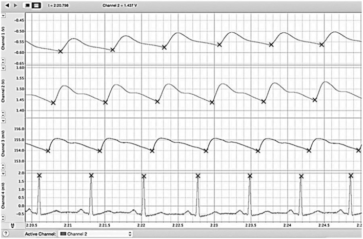
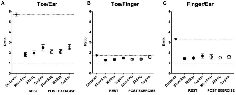
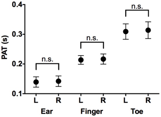
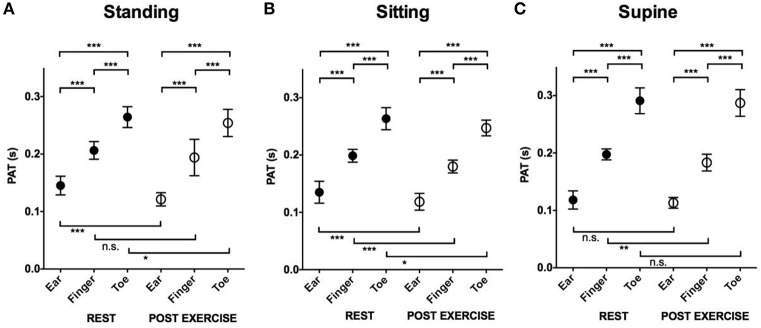
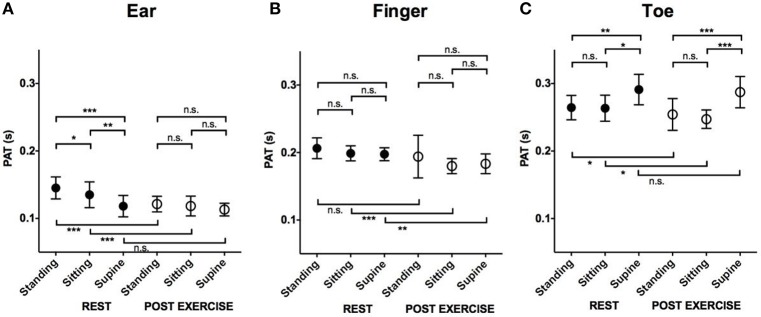
The effects of position and exercise on pulse wave distribution across a healthy, compliant arterial tree are not fully understood. We studied the effects of exercise and position on the pattern of pulse arrival times (PATs) in healthy volunteers. Moreover, we compared the pulse arrival time ratios to the respective distance ratios between different locations. Thirteen young healthy volunteers were studied, using an electrocardiogram and plethysmograph to simultaneously record pulse wave arrival at the ear lobe, index finger and big toe. We compared the differences in PAT between each location at rest and post-exercise in the supine, sitting, and standing position. We also compared the PAT ratio (toe/ear, toe/finger, and finger/ear) to the corresponding pulse path distance ratios. PAT was shortest at the ear then finger and longest at the toe regardless of position or exercise status. PATs were shorter post-exercise compared to rest. When transitioning from a standing to sitting or supine position, PAT to the ear decreased, while the PAT to the toe increased, and PAT to the finger didn't significantly change. PAT ratios were significantly smaller than predicted by the path distance ratios regardless of position or exercise status. Exercise makes PATs shorter. Standing position decrease PAT to the toe and increase to the ear. We conclude that PAT and PAT ratio represent the arterial vascular tree properties as surely as pulse transit time and pulse wave velocity.
姿势和运动对健康、顺应性良好的动脉树中脉搏波分布的影响尚未完全明确。我们研究了运动和姿势对健康志愿者脉搏到达时间(PAT)模式的影响。此外,我们还比较了不同位置之间的脉搏到达时间比与各自的距离比。我们对13名年轻健康志愿者进行了研究,使用心电图和体积描记器同时记录脉搏波到达耳垂、食指和大脚趾的时间。我们比较了仰卧位、坐位和站立位休息时和运动后各位置之间PAT的差异。我们还比较了PAT比(脚趾/耳垂、脚趾/手指和手指/耳垂)与相应的脉搏路径距离比。无论姿势或运动状态如何,PAT在耳垂处最短,在手指处次之,在脚趾处最长。与休息时相比,运动后PAT更短。从站立位转变为坐位或仰卧位时,耳垂处的PAT减小,而脚趾处的PAT增加,手指处的PAT没有显著变化。无论姿势或运动状态如何,PAT比均显著小于路径距离比预测的值。运动使PAT更短。站立位会使脚趾处的PAT减小,耳垂处的PAT增加。我们得出结论,PAT和PAT比与脉搏传播时间和脉搏波速度一样,肯定能代表动脉血管树的特性。