Shi Yu, Dong Kun, Zhang Yu-Guo, Michel René P, Marcus Victoria, Wang Yu-Yue, Chen Yu, Gao Zu-Hua
Department of Pathology, McGill University, Montreal, Quebec QCH3A0G4, Canada.
Department of Laboratory Medicine, Dr. Everett Chalmers Regional Hospital, Horizon Health Network, Fredericton, New Brunswick E3B 5N5, Canada.
World J Gastroenterol. 2017 Feb 7;23(5):792-799. doi: 10.3748/wjg.v23.i5.792.
To investigated the feasibility of using sinusoidal endotheliitis (SE) as a histological marker for liver allograft rejection.
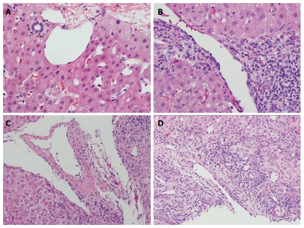
We compared the histological features of 88 liver allograft biopsies with acute cellular rejection (ACR) and 59 cases with no evidence of ACR. SE was scored as: (1) focal linear lifting up of the endothelial cells by lymphocytes with no obvious damage to adjacent hepatocytes; (2) focal disruption of the endothelial lining by a cluster of subendothelial lymphocytes (a group of > 3 lymphocytes); and (3) severe confluent endotheliitis with hemorrhage and adjacent hepatocyte loss.
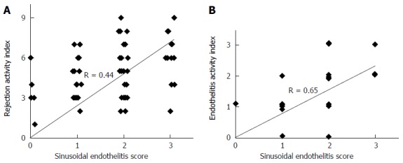
The sensitivity and specificity of SE was 81% and 85%, respectively. Using SE as the only parameter, the positive predictive value for ACR (PPV) was 0.89, whereas the negative predictive value for ACR (NPV) was 0.75. The correlation between RAI and SE was moderate (R = 0.44, < 0.001) (Figure 3A), whereas it became strong (R = 0.65, < 0.001) when correlating SE with the venous endotheliitis activity index only.
Our data suggest that SE scoring could be a reliable and reproducible supplemental parameter to the existing Banff schema for diagnosing acute liver allograft rejection.
探讨将窦状隙内皮炎(SE)作为肝移植排斥反应组织学标志物的可行性。
我们比较了88例有急性细胞排斥反应(ACR)的肝移植活检组织和59例无ACR证据的组织的组织学特征。SE的评分如下:(1)淋巴细胞使内皮细胞局灶性线性抬起,相邻肝细胞无明显损伤;(2)一群内皮下淋巴细胞(一组>3个淋巴细胞)使内皮衬里局灶性破坏;(3)严重融合性内皮炎伴出血及相邻肝细胞丢失。
SE的敏感性和特异性分别为81%和85%。仅将SE作为参数时,ACR的阳性预测值(PPV)为0.89,而ACR的阴性预测值(NPV)为0.75。RAI与SE之间的相关性中等(R = 0.44,P < 0.001)(图3A),而仅将SE与静脉内皮炎活动指数相关时,相关性变强(R = 0.65,P < 0.001)。
我们的数据表明,对于现有的用于诊断急性肝移植排斥反应的Banff标准,SE评分可能是一个可靠且可重复的补充参数。