NeuroImaging Laboratory, University of Pittsburgh, Pittsburgh, Pennsylvania, United States.
UPMC Eye Center, Eye and Ear Institute, Ophthalmology and Visual Science Research Center, Department of Ophthalmology, University of Pittsburgh, Pittsburgh, Pennsylvania, United States.
Sci Rep. 2017 Feb 23;7:43124. doi: 10.1038/srep43124.
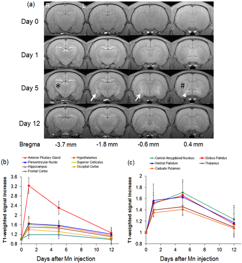
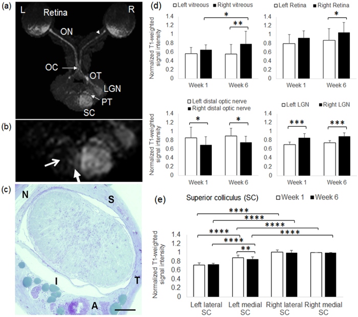
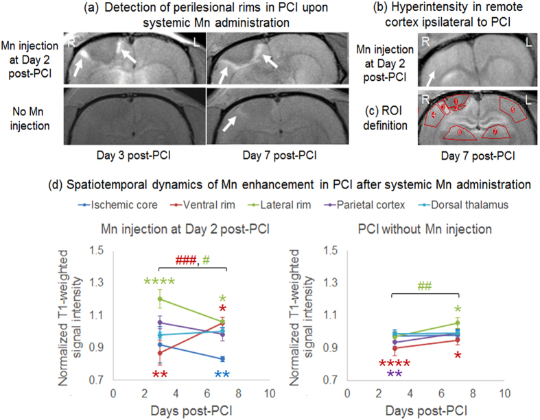
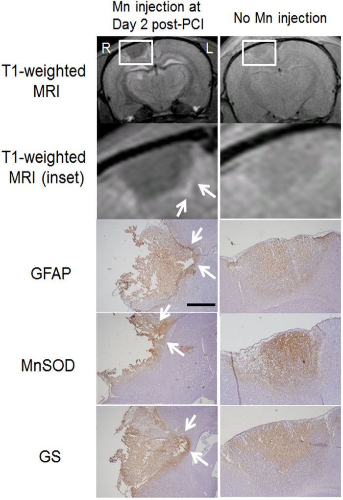
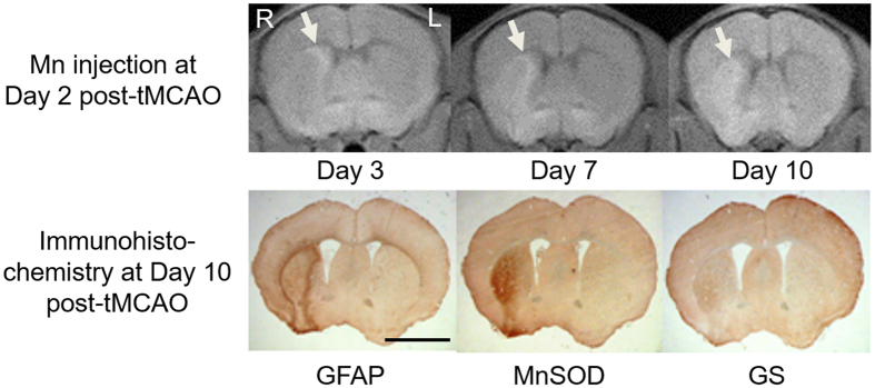
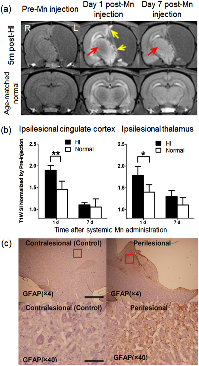
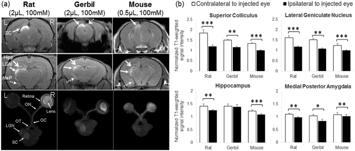
Although manganese (Mn) can enhance brain tissues for improving magnetic resonance imaging (MRI) assessments, the underlying neural mechanisms of Mn detection remain unclear. In this study, we used Mn-enhanced MRI to test the hypothesis that different Mn entry routes and spatiotemporal Mn distributions can reflect different mechanisms of neural circuitry and neurodegeneration in normal and injured brains. Upon systemic administration, exogenous Mn exhibited varying transport rates and continuous redistribution across healthy rodent brain nuclei over a 2-week timeframe, whereas in rodents following photothrombotic cortical injury, transient middle cerebral artery occlusion, or neonatal hypoxic-ischemic brain injury, Mn preferentially accumulated in perilesional tissues expressing gliosis or oxidative stress within days. Intravitreal Mn administration to healthy rodents not only allowed tracing of primary visual pathways, but also enhanced the hippocampus and medial amygdala within a day, whereas partial transection of the optic nerve led to MRI detection of degrading anterograde Mn transport at the primary injury site and the perilesional tissues secondarily over 6 weeks. Taken together, our results indicate the different Mn transport dynamics across widespread projections in normal and diseased brains. Particularly, perilesional brain tissues may attract abnormal Mn accumulation and gradually reduce anterograde Mn transport via specific Mn entry routes.
尽管锰(Mn)可以增强脑组织,从而改善磁共振成像(MRI)评估,但 Mn 检测的潜在神经机制仍不清楚。在这项研究中,我们使用 Mn 增强 MRI 来检验以下假设:不同的 Mn 进入途径和时空 Mn 分布可以反映正常和受损大脑中神经回路和神经退行性变的不同机制。系统给药后,外源性 Mn 在 2 周的时间内以不同的转运速率在健康啮齿动物脑核内连续重新分布,而在光血栓性皮质损伤、短暂性大脑中动脉闭塞或新生儿缺氧缺血性脑损伤后的啮齿动物中,Mn 优先在表达神经胶质增生或氧化应激的病变周围组织中积累。Mn 向健康啮齿动物的玻璃体内给药不仅允许追踪初级视觉通路,而且在一天内还增强了海马体和内侧杏仁核,而视神经的部分横断导致在主要损伤部位和病变周围组织中检测到降解的顺行 Mn 转运,持续 6 周。总之,我们的研究结果表明 Mn 在正常和患病大脑中的广泛投射中具有不同的转运动力学。特别是,病变周围的脑组织可能会吸引异常的 Mn 积累,并通过特定的 Mn 进入途径逐渐减少顺行 Mn 转运。