Haumann Iris, Junghans Dirk, Anstötz Max, Frotscher Michael
Institute of Neuroanatomy, University Medical Center Hamburg-Eppendorf, Hamburg, Germany.
Institute of Embryology and Stem Cell Biology, Department of Biomedicine, University of Basel, Basel, Switzerland.
PLoS One. 2017 Feb 24;12(2):e0172967. doi: 10.1371/journal.pone.0172967. eCollection 2017.
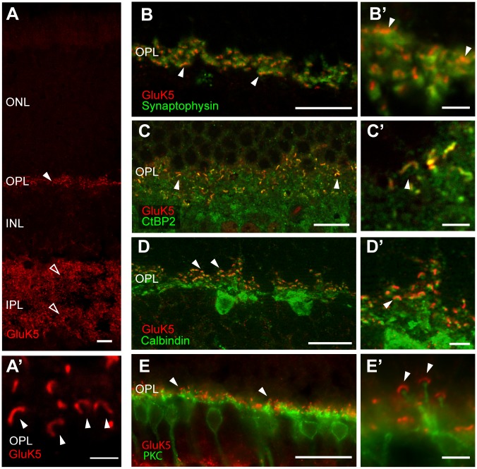
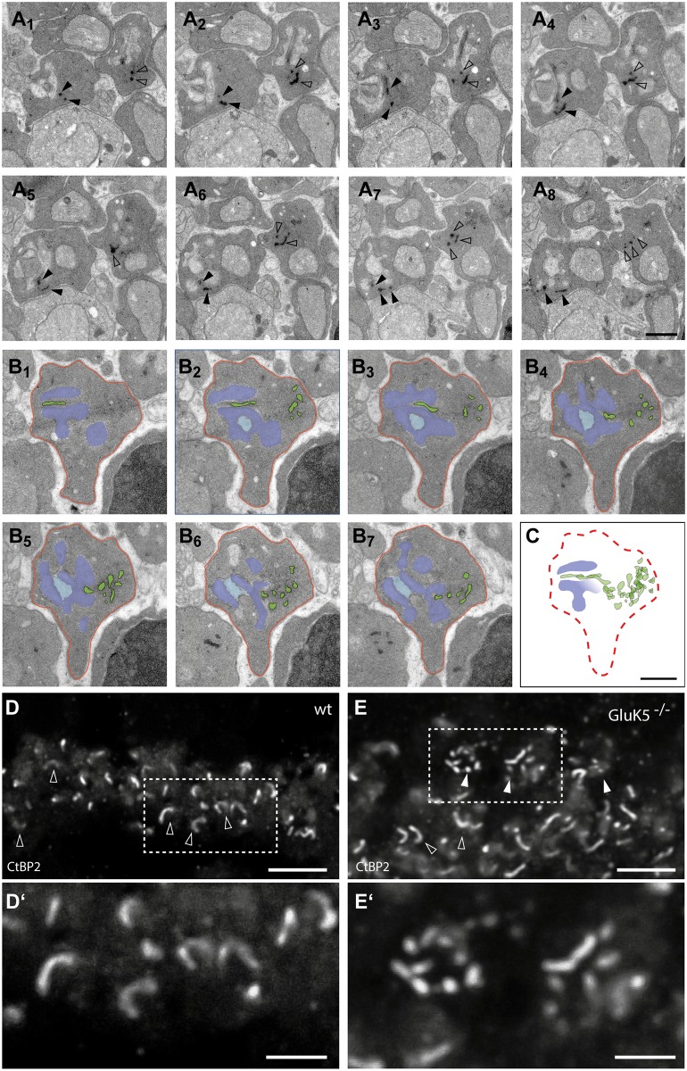
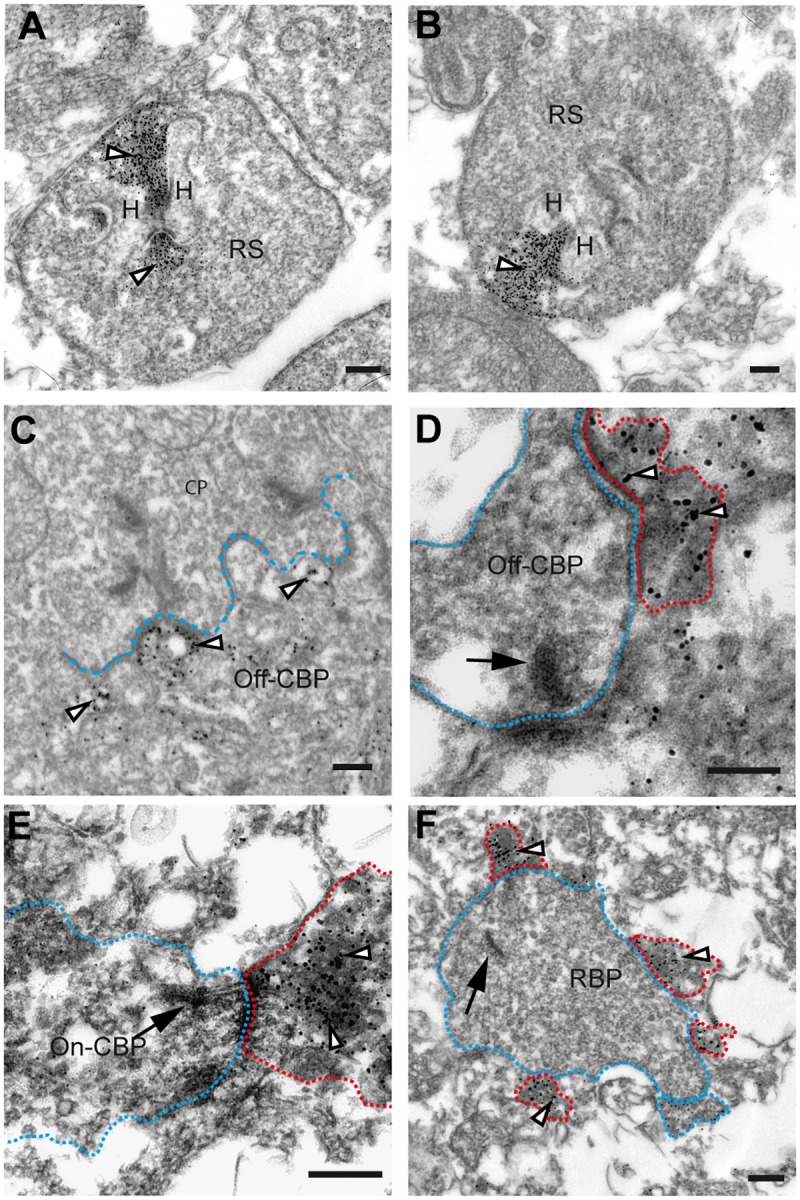
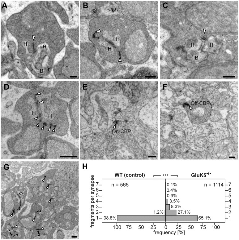
Kainate receptors mediate glutamatergic signaling through both pre- and presynaptic receptors. Here, we studied the expression of the high affinity kainate receptor GluK5 in the mouse retina. Double-immunofluoresence labeling and electron microscopic analysis revealed a presynaptic localization of GluK5 in the outer plexiform layer. Unexpectedly, we found GluK5 almost exclusively localized to the presynaptic ribbon of photoreceptor terminals. Moreover, in GluK5-deficient mutant mice the structural integrity of synaptic ribbons was severely altered pointing to a novel function of GluK5 in organizing synaptic ribbons in the presynaptic terminals of rod photoreceptors.
海人酸受体通过突触前和突触后受体介导谷氨酸能信号传导。在此,我们研究了高亲和力海人酸受体GluK5在小鼠视网膜中的表达。双重免疫荧光标记和电子显微镜分析显示GluK5在外侧网状层的突触前定位。出乎意料的是,我们发现GluK5几乎只定位于光感受器终末的突触前带。此外,在GluK5缺陷型突变小鼠中,突触带的结构完整性严重改变,这表明GluK5在杆状光感受器突触前终末组织突触带方面具有新功能。