• 文献检索
  • 文档翻译
  • 深度研究
  • 学术资讯
  • Suppr Zotero 插件Zotero 插件
  • 邀请有礼
  • 套餐&价格
  • 历史记录
应用&插件
Suppr Zotero 插件Zotero 插件浏览器插件Mac 客户端Windows 客户端微信小程序
定价
高级版会员购买积分包购买API积分包
服务
文献检索文档翻译深度研究API 文档MCP 服务
关于我们
关于 Suppr公司介绍联系我们用户协议隐私条款
关注我们

Suppr 超能文献

核心技术专利:CN118964589B侵权必究
粤ICP备2023148730 号-1Suppr @ 2026

文献检索

告别复杂PubMed语法,用中文像聊天一样搜索,搜遍4000万医学文献。AI智能推荐,让科研检索更轻松。

立即免费搜索

文件翻译

保留排版,准确专业,支持PDF/Word/PPT等文件格式,支持 12+语言互译。

免费翻译文档

深度研究

AI帮你快速写综述,25分钟生成高质量综述,智能提取关键信息,辅助科研写作。

立即免费体验

光学相干断层扫描分析正畸治疗前后的牙釉质厚度

Enamel Thickness before and after Orthodontic Treatment Analysed in Optical Coherence Tomography.

作者信息

Seeliger Julia, Machoy Monika, Koprowski Robert, Safranow Krzysztof, Gedrange Tomasz, Woźniak Krzysztof

机构信息

Division of Orthodontics, Technical University Dresden, Dresden, Germany.

Division of Orthodontics, Pomeranian Medical University, Szczecin, Poland.

出版信息

Biomed Res Int. 2017;2017:8390575. doi: 10.1155/2017/8390575. Epub 2017 Jan 24.

DOI:10.1155/2017/8390575
PMID:28243604
原文链接:https://pmc.ncbi.nlm.nih.gov/articles/PMC5294353/
Abstract

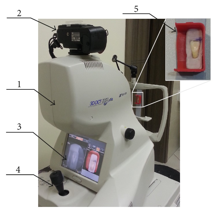
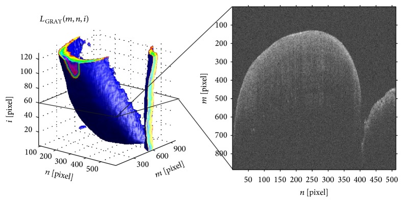
Despite the continuous development of materials and techniques of adhesive bonding, the basic procedure remains relatively constant. The technique is based on three components: etching substance, adhesive system, and composite material. The use of etchants during bonding orthodontic brackets carries the risk of damage to the enamel. Therefore, the article examines the effect of the manner of enamel etching on its thickness before and after orthodontic treatment. The study was carried out in vitro on a group of 80 teeth. It was divided into two subgroups of 40 teeth each. The procedure of enamel etching was performed under laboratory conditions. In the first subgroup, the classic method of enamel etching and the fifth-generation bonding system were used. In the second subgroup, the seventh-generation (self-etching) bonding system was used. In both groups, metal orthodontic brackets were fixed and the enamel was cleaned with a cutter fixed on the micromotor after their removal. Before and after the treatment, two-dimensional optical coherence tomography scans were performed. The enamel thickness was assessed on the two-dimensional scans. The average enamel thickness in both subgroups was not statistically significant.

摘要

尽管粘结技术的材料和工艺不断发展,但其基本程序仍相对固定。该技术基于三个要素:蚀刻剂、粘结系统和复合材料。在粘结正畸托槽时使用蚀刻剂存在损害牙釉质的风险。因此,本文研究了正畸治疗前后牙釉质蚀刻方式对其厚度的影响。该研究在体外对一组80颗牙齿进行。将其分为两个亚组,每组40颗牙齿。牙釉质蚀刻过程在实验室条件下进行。在第一个亚组中,使用经典的牙釉质蚀刻方法和第五代粘结系统。在第二个亚组中,使用第七代(自蚀刻)粘结系统。在两组中,均固定金属正畸托槽,并在拆除托槽后用固定在微型电机上的切割器清洁牙釉质。在治疗前后,进行二维光学相干断层扫描。在二维扫描上评估牙釉质厚度。两个亚组的平均牙釉质厚度无统计学差异。

https://cdn.ncbi.nlm.nih.gov/pmc/blobs/5ce6/5294353/5eae04e5f201/BMRI2017-8390575.002.jpg
https://cdn.ncbi.nlm.nih.gov/pmc/blobs/5ce6/5294353/8233b54bba41/BMRI2017-8390575.001.jpg
https://cdn.ncbi.nlm.nih.gov/pmc/blobs/5ce6/5294353/5eae04e5f201/BMRI2017-8390575.002.jpg
https://cdn.ncbi.nlm.nih.gov/pmc/blobs/5ce6/5294353/8233b54bba41/BMRI2017-8390575.001.jpg
https://cdn.ncbi.nlm.nih.gov/pmc/blobs/5ce6/5294353/5eae04e5f201/BMRI2017-8390575.002.jpg

相似文献

1
Enamel Thickness before and after Orthodontic Treatment Analysed in Optical Coherence Tomography.光学相干断层扫描分析正畸治疗前后的牙釉质厚度
Biomed Res Int. 2017;2017:8390575. doi: 10.1155/2017/8390575. Epub 2017 Jan 24.
2
Optical coherence tomography as a non-invasive method of enamel thickness diagnosis after orthodontic treatment by 3 different types of brackets.光学相干断层扫描作为一种非侵入性方法,用于诊断正畸治疗后三种不同类型托槽对牙釉质厚度的影响。
Adv Clin Exp Med. 2019 Feb;28(2):211-218. doi: 10.17219/acem/79974.
3
Evaluation of changes in enamel thickness after orthodontic treatment depending on the force applied to remove orthodontic brackets: OCT analysis and universal testing machine.根据去除正畸托槽时所施加的力评估正畸治疗后牙釉质厚度的变化:光学相干断层扫描分析和万能材料试验机。
Adv Clin Exp Med. 2019 Jun;28(6):807-813. doi: 10.17219/acem/94141.
4
Automatic method of analysis of OCT images in the assessment of the tooth enamel surface after orthodontic treatment with fixed braces.在固定矫治器正畸治疗后评估牙釉质表面时OCT图像的自动分析方法
Biomed Eng Online. 2014 Apr 22;13:48. doi: 10.1186/1475-925X-13-48.
5
Three-dimensional measurement of residual adhesive and enamel loss on teeth after debonding of orthodontic brackets: an in-vitro study.正畸托槽脱粘后牙齿上残留粘结剂和釉质损失的三维测量:一项体外研究。
Am J Orthod Dentofacial Orthop. 2007 Mar;131(3):301.e9-15. doi: 10.1016/j.ajodo.2006.01.026.
6
Comparison of shear bond strengths of orthodontic brackets bonded to deciduous and permanent teeth.正畸托槽粘结于乳牙和恒牙的剪切粘结强度比较。
Am J Orthod Dentofacial Orthop. 2008 Aug;134(2):198-202. doi: 10.1016/j.ajodo.2006.05.045.
7
Effects of Different Stain Removal Protocols on Bonding Orthodontic Brackets to Enamel.不同脱粘方案对正畸托槽与牙釉质粘结的影响。
J Contemp Dent Pract. 2018 Jul 1;19(7):762-767.
8
Micron-scale human enamel layer characterization after orthodontic bracket debonding by intensity-based layer segmentation in optical coherence tomography images.基于强度的光学相干断层扫描图像分层分割技术对正畸托槽脱落后微米级人牙釉质层的特征分析。
Sci Rep. 2021 May 25;11(1):10831. doi: 10.1038/s41598-021-90354-9.
9
Microleakage under metallic and ceramic brackets bonded with orthodontic self-etching primer systems.使用正畸自酸蚀底漆系统粘结的金属和陶瓷托槽下的微渗漏
Angle Orthod. 2008 Nov;78(6):1089-94. doi: 10.2319/100507-481.1.
10
In vitro Analysis of Shear Bond Strength and Adhesive Remnant Index of Stainless Steel Brackets with Different Adhesive Systems to Enamel.不同粘接系统的不锈钢托槽与牙釉质间剪切粘接强度及粘接剂残留指数的体外分析
J Contemp Dent Pract. 2018 Sep 1;19(9):1047-1051.

引用本文的文献

1
Use of Optical Coherence Tomography in Dentistry.光学相干断层扫描技术在牙科领域的应用。
Int J Dent. 2023 Dec 11;2023:4179210. doi: 10.1155/2023/4179210. eCollection 2023.
2
Shear Bond Strength of Metal and Ceramic Brackets Depending on Etching Protocol in Direct Bonding Technique.直接粘结技术中,金属和陶瓷托槽的剪切粘结强度取决于蚀刻方案
Materials (Basel). 2023 Oct 15;16(20):6697. doi: 10.3390/ma16206697.
3
Comparison of Tooth Color and Enamel and Dentinal Thickness between Orthodontically Treated and Untreated Individuals.

本文引用的文献

1
Automatic method of analysis of OCT images in the assessment of the tooth enamel surface after orthodontic treatment with fixed braces.在固定矫治器正畸治疗后评估牙釉质表面时OCT图像的自动分析方法
Biomed Eng Online. 2014 Apr 22;13:48. doi: 10.1186/1475-925X-13-48.
2
Ultrashort pulsed laser conditioning of human enamel: in vitro study of the influence of geometrical processing parameters on shear bond strength of orthodontic brackets.人牙釉质的超短脉冲激光处理:几何加工参数对正畸托槽剪切粘结强度影响的体外研究
Lasers Med Sci. 2015 Feb;30(2):891-900. doi: 10.1007/s10103-013-1491-2. Epub 2013 Nov 19.
3
Automated analysis of lesion depth and integrated reflectivity in PS-OCT scans of tooth demineralization.
正畸治疗与未治疗个体之间牙齿颜色、牙釉质和牙本质厚度的比较。
Diagnostics (Basel). 2023 Jun 14;13(12):2066. doi: 10.3390/diagnostics13122066.
4
Assessment of the Accuracy in Measuring the Enamel Thickness of Maxillary Incisors with Optical Coherence Tomography.光学相干断层扫描测量上颌切牙牙釉质厚度的准确性评估
Diagnostics (Basel). 2022 Jul 5;12(7):1634. doi: 10.3390/diagnostics12071634.
5
Real-Time Monitoring and Quantitative Evaluation of Resin In-Filtrant Repairing Enamel White Spot Lesions Based on Optical Coherence Tomography.基于光学相干断层扫描技术的树脂浸润剂修复牙釉质白斑病变的实时监测与定量评估
Diagnostics (Basel). 2021 Nov 4;11(11):2046. doi: 10.3390/diagnostics11112046.
6
Transverse Growth of the Maxillo-Mandibular Complex in Untreated Children: A Longitudinal Cone Beam Computed Tomography Study.未治疗儿童的上颌-下颌复合体横向生长:一项纵向锥形束 CT 研究。
Sensors (Basel). 2021 Sep 24;21(19):6378. doi: 10.3390/s21196378.
7
Comparative evaluation of the efficacy of the desensitizing and remineralizing agent in the reduction of dentin hypersensitivity after orthodontic debonding - a randomized clinical trial.正畸去粘结后脱敏再矿化剂降低牙本质过敏疗效的比较评价——一项随机临床试验
Med Pharm Rep. 2021 Apr;94(2):229-238. doi: 10.15386/mpr-1763. Epub 2021 Apr 29.
8
Corrigendum to "Enamel Thickness before and after Orthodontic Treatment Analysed in Optical Coherence Tomography".《光学相干断层扫描分析正畸治疗前后牙釉质厚度》勘误
Biomed Res Int. 2020 May 14;2020:7291314. doi: 10.1155/2020/7291314. eCollection 2020.
9
3-Dimensional characterization of cortical bone microdamage following placement of orthodontic microimplants using Optical Coherence Tomography.使用光学相干断层扫描技术对正畸微种植体植入后皮质骨微损伤的三维特征进行描述。
Sci Rep. 2019 Mar 1;9(1):3242. doi: 10.1038/s41598-019-39670-9.
10
Assessment of cortical bone microdamage following insertion of microimplants using optical coherence tomography: a preliminary study.应用光相干断层扫描评估微种植体植入后皮质骨微损伤:一项初步研究。
J Zhejiang Univ Sci B. 2018;19(11):818-828. doi: 10.1631/jzus.B1700612.
牙齿脱矿偏振敏感光学相干断层扫描(PS-OCT)图像中病损深度和综合反射率的自动分析
Lasers Surg Med. 2010 Jan;42(1):62-8. doi: 10.1002/lsm.20862.
4
Enamel bonding of self-etching and phosphoric acid-etching orthodontic adhesives in simulated clinical conditions: debonding force and enamel surface.模拟临床条件下自酸蚀和磷酸酸蚀正畸粘结剂的釉质粘结:脱粘力与釉质表面
Dent Mater J. 2009 Jul;28(4):419-25. doi: 10.4012/dmj.28.419.
5
Quantification of dental erosions in patients with GERD using optical coherence tomography before and after double-blind, randomized treatment with esomeprazole or placebo.使用光学相干断层扫描技术对胃食管反流病(GERD)患者在接受埃索美拉唑或安慰剂双盲随机治疗前后的牙齿侵蚀情况进行量化分析。
Am J Gastroenterol. 2009 Nov;104(11):2788-95. doi: 10.1038/ajg.2009.441. Epub 2009 Aug 4.
6
Bond strength comparison and scanning electron microscopic evaluation of three orthodontic bonding systems.三种正畸粘结系统的粘结强度比较及扫描电子显微镜评估
Dent Mater J. 2008 May;27(3):392-9. doi: 10.4012/dmj.27.392.
7
Treated enamel surface patterns associated with five orthodontic adhesive systems--surface morphology and shear bond strength.与五种正畸粘结系统相关的处理后牙釉质表面形态及剪切粘结强度
Dent Mater J. 2008 Jan;27(1):1-6. doi: 10.4012/dmj.27.1.
8
Evaluation of failure characteristics and bond strength after ceramic and polycarbonate bracket debonding: effect of bracket base silanization.陶瓷和聚碳酸酯托槽脱粘后的失败特征及粘结强度评估:托槽基底硅烷化的影响
Eur J Orthod. 2008 Apr;30(2):176-82. doi: 10.1093/ejo/cjm100. Epub 2008 Jan 21.
9
Shear bond strength of orthodontic brackets bonded using conventional vs one and two step self-etching/adhesive systems.使用传统方法与一步法和两步法自酸蚀/粘结系统粘结的正畸托槽的剪切粘结强度。
Angle Orthod. 2007 May;77(3):518-23. doi: 10.2319/0003-3219(2007)077[0518:SBSOOB]2.0.CO;2.
10
Scanning electron microscopic evaluation of enamel surfaces exposed to 3 orthodontic bonding systems.对暴露于3种正畸粘结系统的牙釉质表面进行扫描电子显微镜评估。
Am J Orthod Dentofacial Orthop. 2006 Nov;130(5):575-81. doi: 10.1016/j.ajodo.2006.07.002.