Md Noh Mohamad Syafeeq Faeez, Abdul Aziz Ahmad Fuad, Mohd Ghani Khairul Asri, Lee Kheng Siang Christopher, Yunus Rosna, Mohd Yusof Mubarak
Department of Imaging, University Putra Malaysia, Serdang, Malaysia.
Department of Diagnostic Imaging, Hospital Serdang, Serdang, Malaysia.
Am J Case Rep. 2017 Mar 1;18:212-216. doi: 10.12659/ajcr.902101.
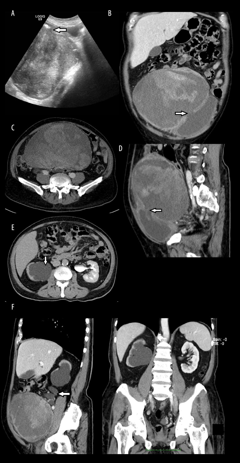
BACKGROUND Intradiverticular bladder tumors are rare. This renders diagnosis of an intradiverticular bladder tumor difficult. Imaging plays a vital role in achieving the diagnosis, and subsequently staging of the disease. CASE REPORT A 74-year-old male presented to our center with a few months history of constitutional symptoms. Upon further history, he reported hematuria two months prior to presentation, which stopped temporarily, only to recur a few days prior to coming to the hospital. The patient admitted to having lower urinary tract symptoms. However, there was no dysuria, no sandy urine, and no fever. Palpation of his abdomen revealed a vague mass at the suprapubic region, which was non tender. In view of his history and the clinical examination findings, an ultrasound of the abdomen and computed tomography (CT) was arranged. These investigations revealed a giant tumor that seemed to be arising from a bladder diverticulum, with a mass effect and hydronephrosis. He later underwent operative intervention. CONCLUSIONS Intradiverticular bladder tumors may present a challenge to the treating physician in an atypical presentation; thus requiring a high index of suspicion and knowledge of tumor pathophysiology. As illustrated in our case, CT with its wide availability and multiplanar imaging capabilities offers a useful means for diagnosis, disease staging, operative planning, and follow-up.
背景 膀胱憩室内肿瘤罕见。这使得膀胱憩室内肿瘤的诊断具有挑战性。影像学检查在实现诊断及随后的疾病分期中起着至关重要的作用。病例报告 一名74岁男性因有几个月的全身症状病史前来我院就诊。进一步询问病史时,他报告在就诊前两个月出现血尿,血尿曾暂时停止,但在入院前几天又复发。患者承认有下尿路症状。然而,无尿痛、无沙粒样尿、无发热。触诊其腹部发现耻骨上区有一模糊肿块,无压痛。鉴于其病史及临床检查结果,安排了腹部超声和计算机断层扫描(CT)检查。这些检查发现一个巨大肿瘤,似乎起源于膀胱憩室,伴有占位效应和肾积水。他随后接受了手术干预。结论 膀胱憩室内肿瘤可能以非典型表现给治疗医生带来挑战;因此需要高度的怀疑指数和对肿瘤病理生理学的了解。如我们的病例所示,CT因其广泛可用性和多平面成像能力,为诊断、疾病分期、手术规划及随访提供了一种有用的手段。