Shen Guohua, Lan You, Zhang Kan, Ren Pengwei, Jia Zhiyun
Department of Nuclear Medicine, West China Hospital, Sichuan University, Chengdu, Sichuan, People's Republic of China.
Division of Laboratory Medicine, West China School of Medicine, Sichuan University, Chengdu, Sichuan, People's Republic of China.
PLoS One. 2017 Mar 2;12(3):e0173104. doi: 10.1371/journal.pone.0173104. eCollection 2017.
Accurate clinical staging of mediastinal lymph nodes of patients with lung cancer is important in determining therapeutic options and prognoses. We aimed to compare the diagnostic performance of diffusion-weighted magnetic resonance imaging (DWI) and 18F-fluorodeoxyglucose positron emission tomography/computed tomography (18F-FDG PET/CT) in detecting mediastinal nodal metastasis of lung cancer.
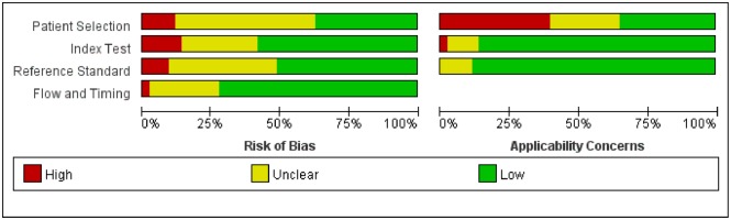
Relevant studies were systematically searched in the MEDLINE, EMBASE, PUBMED, and Cochrane Library databases. Based on extracted data, the pooled sensitivity, specificity, positive and negative likelihood ratios (PLR and NLR) with individual 95% confidence intervals were calculated. In addition, the publication bias was assessed by Deek's funnel plot of the asymmetry test. The potential heterogeneity was explored by threshold effect analysis and subgroup analyses.
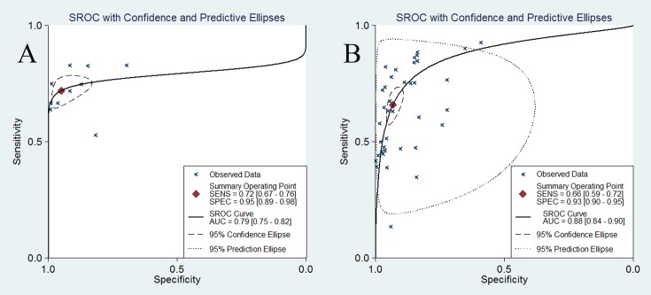
Forty-three studies were finally included. For PET/CT, the pooled sensitivity and specificity were 0.65 (0.63-0.67) and 0.93 (0.93-0.94), respectively. The corresponding values of DWI were 0.72 (0.68-0.76) and 0.97 (0.96-0.98), respectively. The overall PLR and NLR of DWI were 13.15 (5.98-28.89) and 0.32 (0.27-0.39), respectively. For PET/CT, the corresponding values were 8.46 (6.54-10.96) and 0.38 (0.33-0.45), respectively. The Deek's test revealed no significant publication bias. Study design and patient enrollment were potential causes for the heterogeneity of DWI studies and the threshold was a potential source for PET/CT studies.
Both modalities are beneficial in detecting lymph nodes metastases in lung cancer without significant differences between them. DWI might be an alternative modality for evaluating nodal status of NSCLC.
准确对肺癌患者纵隔淋巴结进行临床分期对于确定治疗方案和预后至关重要。我们旨在比较扩散加权磁共振成像(DWI)和18F-氟脱氧葡萄糖正电子发射断层扫描/计算机断层扫描(18F-FDG PET/CT)在检测肺癌纵隔淋巴结转移方面的诊断性能。
在MEDLINE、EMBASE、PUBMED和Cochrane图书馆数据库中系统检索相关研究。根据提取的数据,计算合并敏感度、特异度、阳性和阴性似然比(PLR和NLR)及各自的95%置信区间。此外,通过Deek不对称检验的漏斗图评估发表偏倚。通过阈值效应分析和亚组分析探讨潜在的异质性。
最终纳入43项研究。对于PET/CT,合并敏感度和特异度分别为0.65(0.63 - 0.67)和0.93(0.93 - 0.94)。DWI的相应值分别为0.72(0.68 - 0.76)和0.97(0.96 - 0.98)。DWI的总体PLR和NLR分别为13.15(5.98 - 28.89)和0.32(0.27 - 0.39)。对于PET/CT,相应值分别为8.46(6.54 - 10.96)和0.38(0.33 - 0.45)。Deek检验显示无显著发表偏倚。研究设计和患者纳入是DWI研究异质性的潜在原因,而阈值是PET/CT研究的潜在异质性来源。
两种方法在检测肺癌淋巴结转移方面均有益,且两者之间无显著差异。DWI可能是评估非小细胞肺癌淋巴结状态的替代方法。