• 文献检索
  • 文档翻译
  • 深度研究
  • 学术资讯
  • Suppr Zotero 插件Zotero 插件
  • 邀请有礼
  • 套餐&价格
  • 历史记录
应用&插件
Suppr Zotero 插件Zotero 插件浏览器插件Mac 客户端Windows 客户端微信小程序
定价
高级版会员购买积分包购买API积分包
服务
文献检索文档翻译深度研究API 文档MCP 服务
关于我们
关于 Suppr公司介绍联系我们用户协议隐私条款
关注我们

Suppr 超能文献

核心技术专利:CN118964589B侵权必究
粤ICP备2023148730 号-1Suppr @ 2026

文献检索

告别复杂PubMed语法,用中文像聊天一样搜索,搜遍4000万医学文献。AI智能推荐,让科研检索更轻松。

立即免费搜索

文件翻译

保留排版,准确专业,支持PDF/Word/PPT等文件格式,支持 12+语言互译。

免费翻译文档

深度研究

AI帮你快速写综述,25分钟生成高质量综述,智能提取关键信息,辅助科研写作。

立即免费体验

在多次样品施加和印迹后进行玻璃化处理可提高冷冻电子显微镜网格上的颗粒密度。

Vitrification after multiple rounds of sample application and blotting improves particle density on cryo-electron microscopy grids.

作者信息

Snijder Joost, Borst Andrew J, Dosey Annie, Walls Alexandra C, Burrell Anika, Reddy Vijay S, Kollman Justin M, Veesler David

机构信息

Department of Biochemistry, University of Washington, Seattle, WA, USA.

Department of Integrative Computational and Structural Biology, The Scripps Research Institute, La Jolla, CA, USA.

出版信息

J Struct Biol. 2017 Apr;198(1):38-42. doi: 10.1016/j.jsb.2017.02.008. Epub 2017 Feb 22.

DOI:10.1016/j.jsb.2017.02.008
PMID:28254381
原文链接:https://pmc.ncbi.nlm.nih.gov/articles/PMC5400742/
Abstract

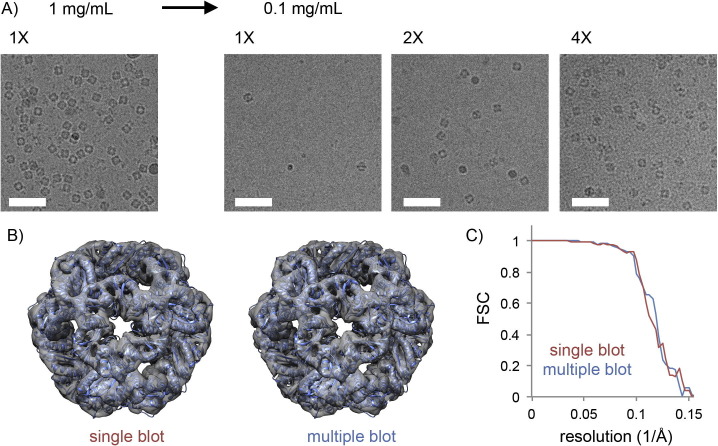
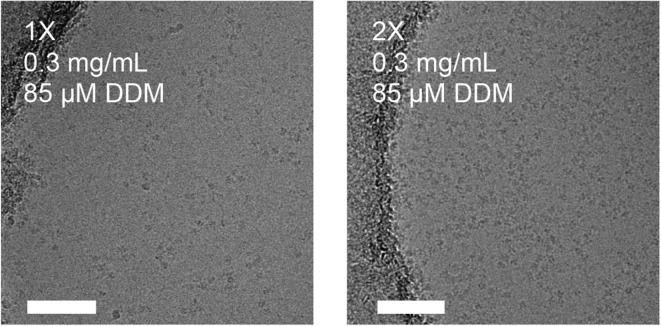
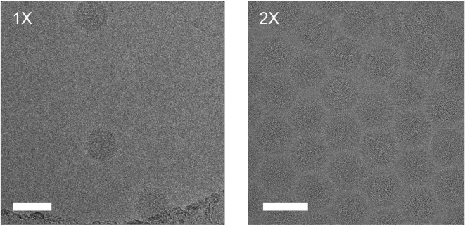
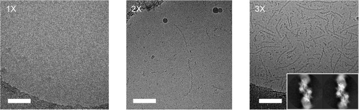
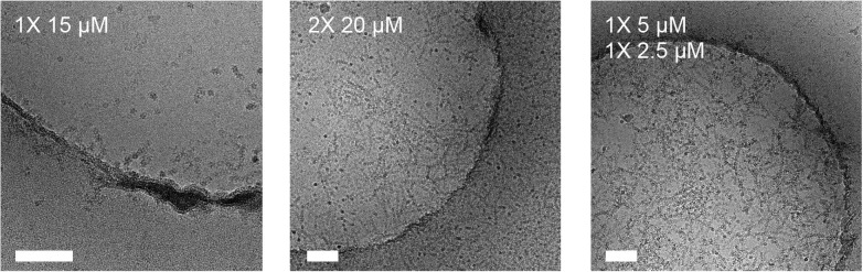
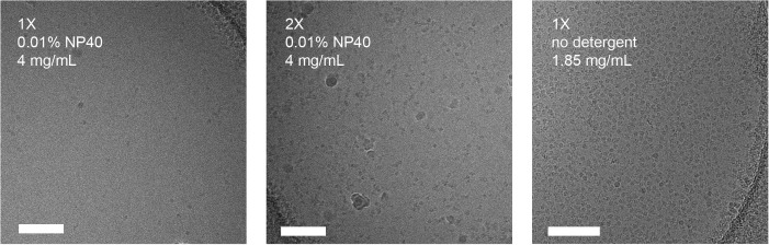
Single particle cryo-electron microscopy (cryoEM) is becoming widely adopted as a tool for structural characterization of biomolecules at near-atomic resolution. Vitrification of the sample to obtain a dense distribution of particles within a single field of view remains a major bottleneck for the success of such experiments. Here, we describe a simple and cost-effective method to increase the density of frozen-hydrated particles on grids with holey carbon support films. It relies on performing multiple rounds of sample application and blotting prior to plunge freezing in liquid ethane. We show that this approach is generally applicable and significantly increases particle density for a range of samples, such as small protein complexes, viruses and filamentous assemblies. The method is versatile, easy to implement, minimizes sample requirements and can enable characterization of samples that would otherwise resist structural studies using single particle cryoEM.

摘要

单颗粒冷冻电子显微镜技术(cryoEM)正被广泛用作一种在近原子分辨率下对生物分子进行结构表征的工具。将样品玻璃化以在单个视野内获得密集的颗粒分布仍然是此类实验成功的主要瓶颈。在此,我们描述了一种简单且经济高效的方法,用于提高带有多孔碳支撑膜的网格上冷冻水合颗粒的密度。该方法依赖于在液氮中快速冷冻之前进行多轮样品施加和吸印操作。我们表明,这种方法普遍适用,并且能显著提高一系列样品(如小蛋白质复合物、病毒和丝状聚集体)的颗粒密度。该方法用途广泛、易于实施,将样品需求降至最低,并且能够对那些否则将无法使用单颗粒冷冻电子显微镜技术进行结构研究的样品进行表征。

https://cdn.ncbi.nlm.nih.gov/pmc/blobs/d412/7119143/c6c9e3925fd6/gr6_lrg.jpg
https://cdn.ncbi.nlm.nih.gov/pmc/blobs/d412/7119143/888f2e2cf5ab/gr1_lrg.jpg
https://cdn.ncbi.nlm.nih.gov/pmc/blobs/d412/7119143/4cdbad053e6a/gr2_lrg.jpg
https://cdn.ncbi.nlm.nih.gov/pmc/blobs/d412/7119143/ed0845a85215/gr3_lrg.jpg
https://cdn.ncbi.nlm.nih.gov/pmc/blobs/d412/7119143/bdacbf551730/gr4_lrg.jpg
https://cdn.ncbi.nlm.nih.gov/pmc/blobs/d412/7119143/75aeeb48841c/gr5_lrg.jpg
https://cdn.ncbi.nlm.nih.gov/pmc/blobs/d412/7119143/c6c9e3925fd6/gr6_lrg.jpg

相似文献

1
Vitrification after multiple rounds of sample application and blotting improves particle density on cryo-electron microscopy grids.在多次样品施加和印迹后进行玻璃化处理可提高冷冻电子显微镜网格上的颗粒密度。
J Struct Biol. 2017 Apr;198(1):38-42. doi: 10.1016/j.jsb.2017.02.008. Epub 2017 Feb 22.
2
Spotiton: a prototype for an integrated inkjet dispense and vitrification system for cryo-TEM.Spotiton:用于 cryo-TEM 的集成喷墨分配和玻璃化系统的原型。
J Struct Biol. 2012 Jul;179(1):68-75. doi: 10.1016/j.jsb.2012.04.020. Epub 2012 May 5.
3
Preparation of macromolecular complexes for cryo-electron microscopy.用于冷冻电子显微镜的大分子复合物的制备。
Nat Protoc. 2007;2(12):3239-46. doi: 10.1038/nprot.2007.452.
4
Manual Blot-and-Plunge Freezing of Biological Specimens for Single-Particle Cryogenic Electron Microscopy.手动印迹-浸入式生物标本冷冻技术用于单颗粒低温电子显微镜。
J Vis Exp. 2022 Feb 7(180). doi: 10.3791/62765.
5
Optimizing "self-wicking" nanowire grids.优化“自吸水”纳米线网格。
J Struct Biol. 2018 May;202(2):170-174. doi: 10.1016/j.jsb.2018.01.001. Epub 2018 Jan 6.
6
A method to achieve homogeneous dispersion of large transmembrane complexes within the holes of carbon films for electron cryomicroscopy.一种实现大跨膜复合物在碳膜孔内均匀分散的方法,用于电子冷冻显微镜。
J Struct Biol. 2013 Apr;182(1):51-6. doi: 10.1016/j.jsb.2013.01.004. Epub 2013 Jan 26.
7
Preparation of Proteins and Macromolecular Assemblies for Cryo-electron Microscopy.用于冷冻电子显微镜的蛋白质和大分子组装体的制备
Methods Mol Biol. 2020;2073:221-246. doi: 10.1007/978-1-4939-9869-2_13.
8
Direct transfer of electron microscopy samples to wetted carbon and graphene films via a support floatation block.通过支撑漂浮块将电子显微镜样品直接转移到湿润的碳和石墨烯薄膜上。
J Struct Biol. 2021 Mar;213(1):107677. doi: 10.1016/j.jsb.2020.107677. Epub 2020 Dec 8.
9
Do's and don'ts of cryo-electron microscopy: a primer on sample preparation and high quality data collection for macromolecular 3D reconstruction.冷冻电子显微镜的注意事项:大分子三维重建的样品制备和高质量数据收集入门指南。
J Vis Exp. 2015 Jan 9(95):52311. doi: 10.3791/52311.
10
Preparing Frozen-Hydrated Protein-Nucleic Acid Assemblies for High-Resolution Cryo-EM Imaging.制备用于高分辨率冷冻电镜成像的冷冻水合蛋白质-核酸组装体。
Methods Mol Biol. 2018;1814:287-296. doi: 10.1007/978-1-4939-8591-3_17.

引用本文的文献

1
Collagen VI microfibril structure reveals mechanism for molecular assembly and clustering of inherited pathogenic mutations.胶原蛋白VI微原纤维结构揭示了遗传性致病突变的分子组装和聚集机制。
Nat Commun. 2025 Aug 14;16(1):7549. doi: 10.1038/s41467-025-62923-3.
2
Passage of ribosomes through microsprayer increases functional activity - Implications for activity assays in time-resolved cryo-EM.核糖体通过微喷雾器会增加功能活性——对时间分辨冷冻电镜中活性测定的启示。
J Struct Biol. 2025 Sep;217(3):108232. doi: 10.1016/j.jsb.2025.108232. Epub 2025 Jul 4.
3
Microfluidics combined with electron microscopy for rapid and high-throughput mapping of antibody-viral glycoprotein complexes.

本文引用的文献

1
Cryo-EM structure of haemoglobin at 3.2 Å determined with the Volta phase plate.采用 Volta 相板技术获得的 3.2 Å 分辨率血影蛋白冷冻电镜结构。
Nat Commun. 2017 Jun 30;8:16099. doi: 10.1038/ncomms16099.
2
MotionCor2: anisotropic correction of beam-induced motion for improved cryo-electron microscopy.MotionCor2:用于改进冷冻电子显微镜的束流诱导运动的各向异性校正
Nat Methods. 2017 Apr;14(4):331-332. doi: 10.1038/nmeth.4193. Epub 2017 Feb 27.
3
Antibody-Based Affinity Cryoelectron Microscopy at 2.6-Å Resolution.基于抗体的亲和冷冻电子显微镜技术,分辨率达2.6埃
微流控技术与电子显微镜相结合,用于抗体-病毒糖蛋白复合物的快速高通量图谱绘制。
Nat Biomed Eng. 2025 Jun 3. doi: 10.1038/s41551-025-01411-x.
4
Structure of bifunctional variediene synthase yields unique insight on biosynthetic diterpene assembly and cyclization.双功能多烯合酶的结构为二萜生物合成组装和环化提供了独特见解。
Nat Commun. 2025 Jun 2;16(1):5089. doi: 10.1038/s41467-025-60537-3.
5
Structure-based validation of recombinant light-harvesting complex II.基于结构的重组捕光复合物II的验证
PNAS Nexus. 2024 Sep 25;3(9):pgae405. doi: 10.1093/pnasnexus/pgae405. eCollection 2024 Sep.
6
Small LEA proteins mitigate air-water interface damage to fragile cryo-EM samples during plunge freezing.小分子 LEA 蛋白可减轻在 plunge 冷冻过程中脆弱的 cryo-EM 样品在气-液界面处的损伤。
Nat Commun. 2024 Sep 4;15(1):7705. doi: 10.1038/s41467-024-52091-1.
7
Self-assembled superstructure alleviates air-water interface effect in cryo-EM.自组装超结构减轻了 cryo-EM 中的气-液界面效应。
Nat Commun. 2024 Aug 24;15(1):7300. doi: 10.1038/s41467-024-51696-w.
8
encapsulin cargo protein EncD is a flavin-binding protein with ferric reductase activity.被囊蛋白货物蛋白 EncD 是一种黄素结合蛋白,具有铁还原酶活性。
Proc Natl Acad Sci U S A. 2024 May 21;121(21):e2400426121. doi: 10.1073/pnas.2400426121. Epub 2024 May 15.
9
Structure of the γ-tubulin ring complex-capped microtubule.γ-微管蛋白环复合物封端的微管结构。
Nat Struct Mol Biol. 2024 Jul;31(7):1124-1133. doi: 10.1038/s41594-024-01264-z. Epub 2024 Apr 12.
10
Structure of full-length ERGIC-53 in complex with MCFD2 for cargo transport.全长 ERGIC-53 与 MCFD2 复合物的结构用于货物运输。
Nat Commun. 2024 Mar 16;15(1):2404. doi: 10.1038/s41467-024-46747-1.
Structure. 2016 Nov 1;24(11):1984-1990. doi: 10.1016/j.str.2016.09.008.
4
Crucial steps in the structure determination of a coronavirus spike glycoprotein using cryo-electron microscopy.使用冷冻电子显微镜确定冠状病毒刺突糖蛋白结构的关键步骤。
Protein Sci. 2017 Jan;26(1):113-121. doi: 10.1002/pro.3048. Epub 2016 Oct 18.
5
Glycan shield and epitope masking of a coronavirus spike protein observed by cryo-electron microscopy.通过冷冻电子显微镜观察到的冠状病毒刺突蛋白的聚糖屏蔽和表位掩盖
Nat Struct Mol Biol. 2016 Oct;23(10):899-905. doi: 10.1038/nsmb.3293. Epub 2016 Sep 12.
6
Cryo-electron microscopy structure of a coronavirus spike glycoprotein trimer.冠状病毒刺突糖蛋白三聚体的冷冻电子显微镜结构
Nature. 2016 Mar 3;531(7592):114-117. doi: 10.1038/nature16988. Epub 2016 Feb 8.
7
An electron microscopy journey in the study of microtubule structure and dynamics.微管结构与动力学研究中的电子显微镜之旅。
Protein Sci. 2015 Dec;24(12):1912-9. doi: 10.1002/pro.2808. Epub 2015 Oct 11.
8
Cryo-EM: A Unique Tool for the Visualization of Macromolecular Complexity.冷冻电镜:一种用于可视化大分子复杂性的独特工具。
Mol Cell. 2015 May 21;58(4):677-89. doi: 10.1016/j.molcel.2015.02.019.
9
A primer to single-particle cryo-electron microscopy.单颗粒冷冻电子显微镜入门
Cell. 2015 Apr 23;161(3):438-449. doi: 10.1016/j.cell.2015.03.050.
10
2.8 Å resolution reconstruction of the Thermoplasma acidophilum 20S proteasome using cryo-electron microscopy.利用冷冻电子显微镜对嗜热栖热菌20S蛋白酶体进行2.8埃分辨率的重建。
Elife. 2015 Mar 11;4:e06380. doi: 10.7554/eLife.06380.