Buckle Tessa, van der Wal Steffen, van Malderen Stijn J M, Müller Larissa, Kuil Joeri, van Unen Vincent, Peters Ruud J B, van Bemmel Margaretha E M, McDonnell Liam A, Velders Aldrik H, Koning Frits, Vanhaeke Frank, van Leeuwen Fijs W B
Interventional Molecular Imaging laboratory, Department of Radiology, Leiden University Medical Center, Leiden, the Netherlands;; Division of Molecular Pathology, Netherlands Cancer Institute- Antoni van Leeuwenhoek hospital (NKI-AvL), Amsterdam, the Netherlands.
Interventional Molecular Imaging laboratory, Department of Radiology, Leiden University Medical Center, Leiden, the Netherlands.
Theranostics. 2017 Jan 12;7(3):624-633. doi: 10.7150/thno.17484. eCollection 2017.
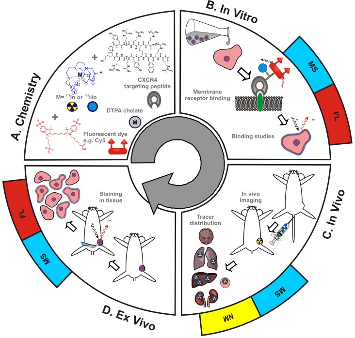
Development of theranostic concepts that include inductively coupled plasma mass spectrometry (ICP-MS) and laser ablation ICP-MS (LA-ICP-MS) imaging can be hindered by the lack of a direct comparison to more standardly used methods for and evaluation; e.g. fluorescence or nuclear medicine. In this study a bimodal (or rather, hybrid) tracer that contains both a fluorescent dye and a chelate was used to evaluate the existence of a direct link between mass spectrometry (MS) and and molecular imaging findings using fluorescence and radioisotopes. At the same time, the hybrid label was used to determine whether the use of a single isotope label would allow for MS-based diagnostics.
A hybrid label that contained both a DTPA chelate (that was coordinated with either Ho or In) and a Cy5 fluorescent dye was coupled to the chemokine receptor 4 (CXCR4) targeting peptide Ac-TZ14011 (hybrid-Cy5-Ac-TZ4011). This receptor targeting tracer was used to 1) validate the efficacy of (Ho-based) mass-cytometry in determining the receptor affinity via comparison with fluorescence-based flow cytometry (Cy5), 2) evaluate the microscopic binding pattern of the tracer in tumor cells using both fluorescence confocal imaging (Cy5) and LA-ICP-MS-imaging (Ho), 3) compare biodistribution patterns obtained with ICP-MS (Ho) and radiodetection (In) after intravenous administration of hybrid-Cy5-Ac-TZ4011 in tumor-bearing mice. Finally, LA-ICP-MS-imaging (Ho) was linked to fluorescence-based analysis of excised tissue samples (Cy5).
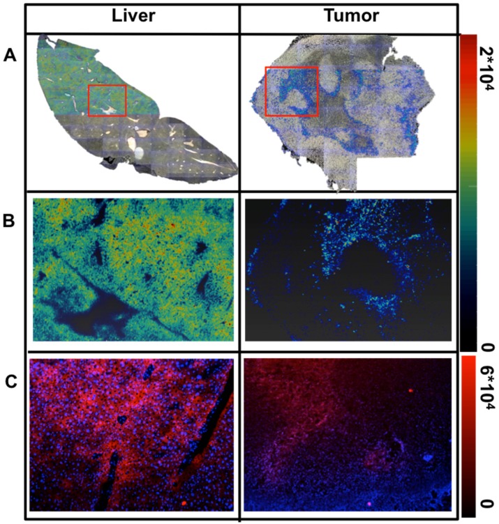
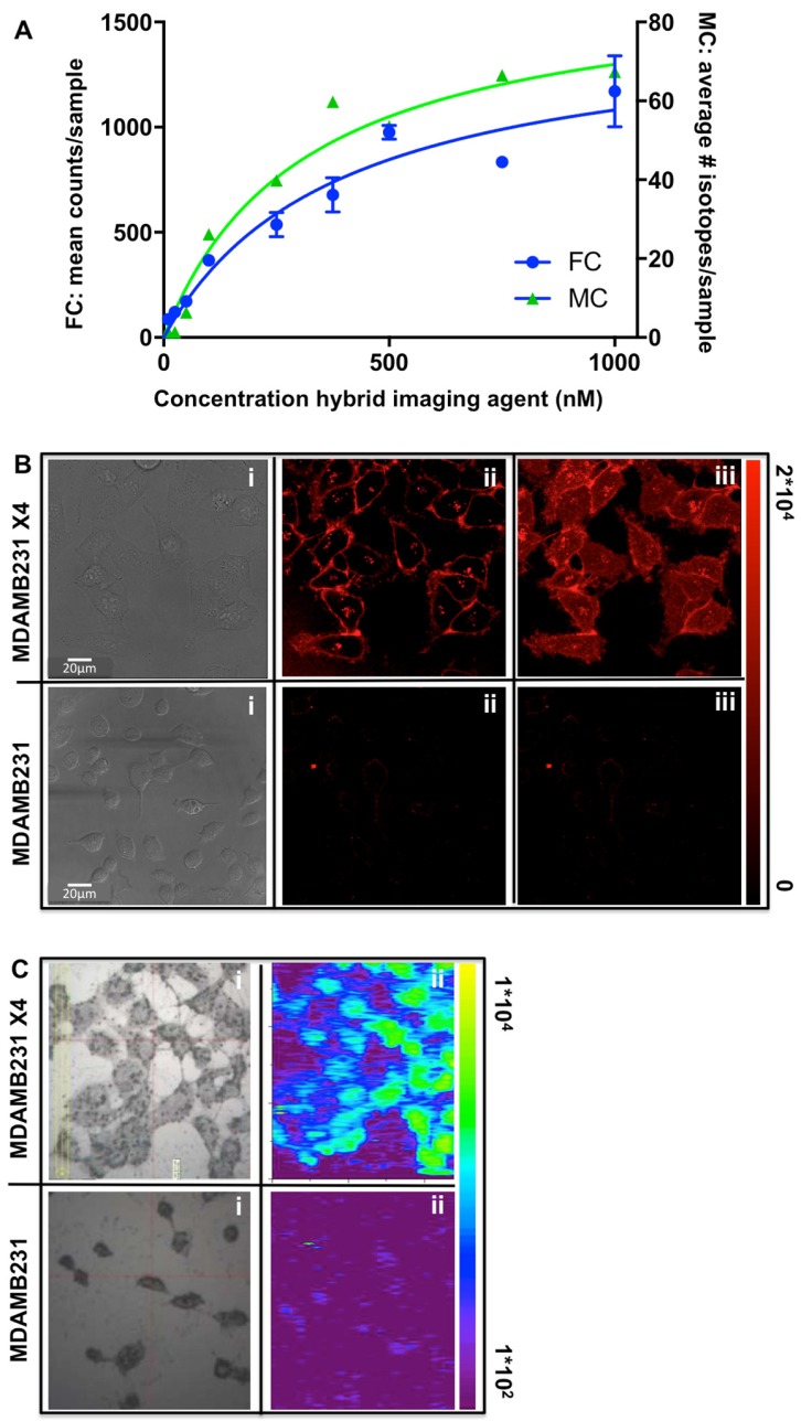
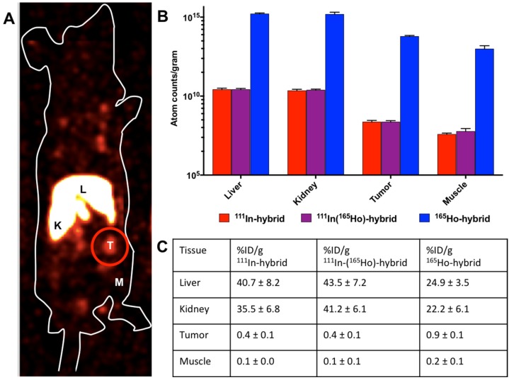
Analysis with both mass-cytometry and flow cytometry revealed a similar receptor affinity, respectively 352 ± 141 nM and 245 ± 65 nM (p = 0.08), but with a much lower detection sensitivity for the first modality. LA-ICP-MS imaging (Ho) enabled clear discrimination between CXCR4 positive and negative cells, but fluorescence microscopy was required to determine the intracellular distribution. biodistribution patterns obtained with ICP-MS (Ho) and radiodetection (In) of the hybrid peptide were shown to be similar. Assessment of tracer distribution in excised tissues revealed the location of tracer uptake with both LA-ICP-MS-imaging and fluorescence imaging.
Lanthanide-isotope chelation expands the scope of fluorescent/radioactive hybrid tracers to include MS-based analytical tools such as mass-cytometry, ICP-MS and LA-ICP-MS imaging in molecular pathology. In contradiction to common expectations, MS detection using a single chelate imaging agent was shown to be feasible, enabling a direct link between nuclear medicine-based imaging and theranostic methods.
包括电感耦合等离子体质谱(ICP-MS)和激光烧蚀ICP-MS(LA-ICP-MS)成像在内的诊疗一体化概念的发展可能会受到阻碍,因为缺乏与更常用的评估方法(例如荧光或核医学方法)的直接比较。在本研究中,使用一种同时包含荧光染料和螯合物的双峰(或者更确切地说是混合)示踪剂,来评估质谱(MS)与使用荧光和放射性同位素的分子成像结果之间是否存在直接联系。同时,该混合标记物用于确定使用单一同位素标记是否能实现基于MS的诊断。
一种同时包含二乙三胺五乙酸(DTPA)螯合物(与钬或铟配位)和Cy5荧光染料的混合标记物与趋化因子受体4(CXCR4)靶向肽Ac-TZ14011偶联(混合-Cy5-Ac-TZ4011)。这种受体靶向示踪剂用于:1)通过与基于荧光的流式细胞术(Cy5)比较,验证(基于钬的)质谱细胞术在确定受体亲和力方面的有效性;2)使用荧光共聚焦成像(Cy5)和LA-ICP-MS成像(钬)评估示踪剂在肿瘤细胞中的微观结合模式;3)在荷瘤小鼠静脉注射混合-Cy5-Ac-TZ4011后,比较通过ICP-MS(钬)和放射性检测(铟)获得的生物分布模式。最后,将LA-ICP-MS成像(钬)与切除组织样本的基于荧光的分析(Cy5)联系起来。
质谱细胞术和流式细胞术分析分别显示出相似的受体亲和力,分别为352±141 nM和245±65 nM(p = 0.08),但第一种方法的检测灵敏度要低得多。LA-ICP-MS成像(钬)能够清晰区分CXCR4阳性和阴性细胞,但需要荧光显微镜来确定细胞内分布。混合肽通过ICP-MS(钬)和放射性检测(铟)获得的生物分布模式显示相似。对切除组织中示踪剂分布的评估揭示了通过LA-ICP-MS成像和荧光成像摄取示踪剂的位置。
镧系元素同位素螯合扩展了荧光/放射性混合示踪剂的范围,将基于MS的分析工具(如质谱细胞术、ICP-MS和LA-ICP-MS成像)纳入分子病理学。与普遍预期相反,使用单一螯合成像剂进行MS检测被证明是可行的,实现了基于核医学的成像与诊疗一体化方法之间的直接联系。