Jia Yan-Bo, Jiang Dian-Ming, Ren Yi-Zhong, Liang Zi-Hong, Zhao Zhen-Qun, Wang Yu-Xin
Department of Orthopaedics, The First Affiliated Hospital of Chongqing Medical University, Yuanjia Gang, Chongqing 400016, P.R. China.
Department of Orthopaedics, The Second Affiliated Hospital of Inner Mongolia Medical University, Huhhot, Inner Mongolia 010030, P.R. China.
Mol Med Rep. 2017 Apr;15(4):1585-1592. doi: 10.3892/mmr.2017.6160. Epub 2017 Feb 2.
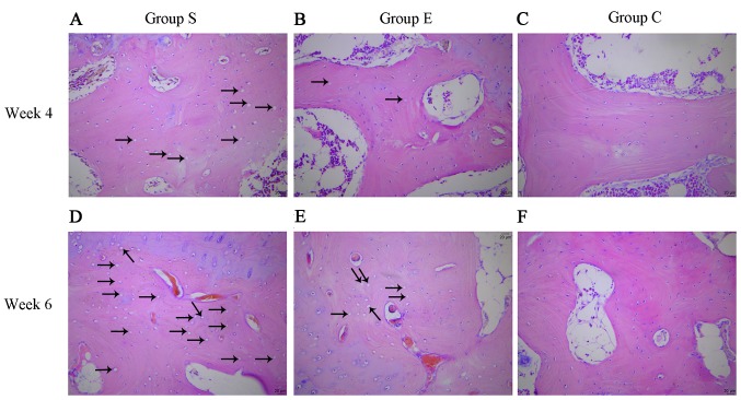
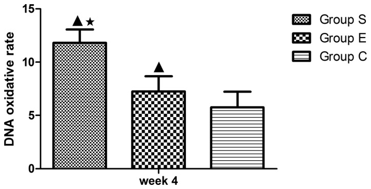
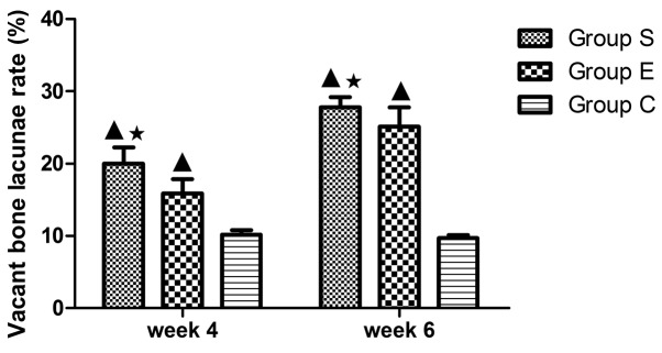
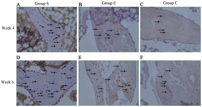
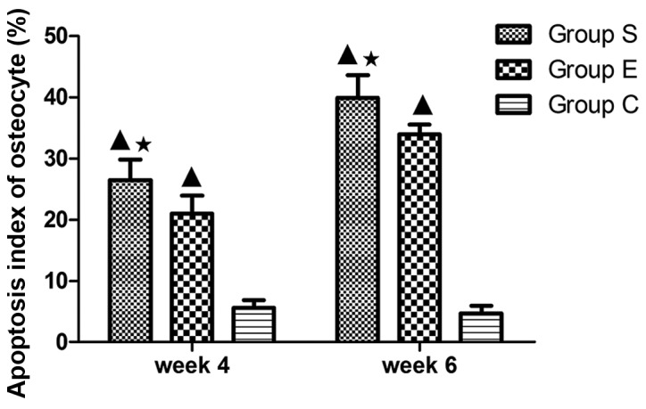
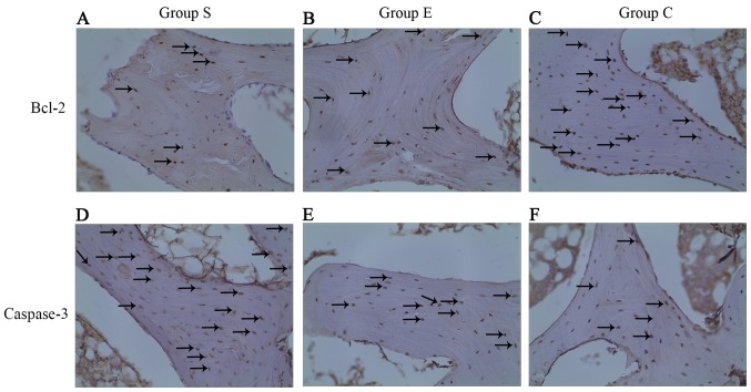
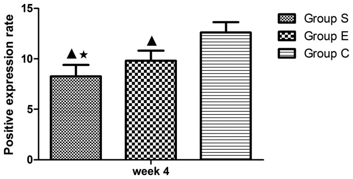
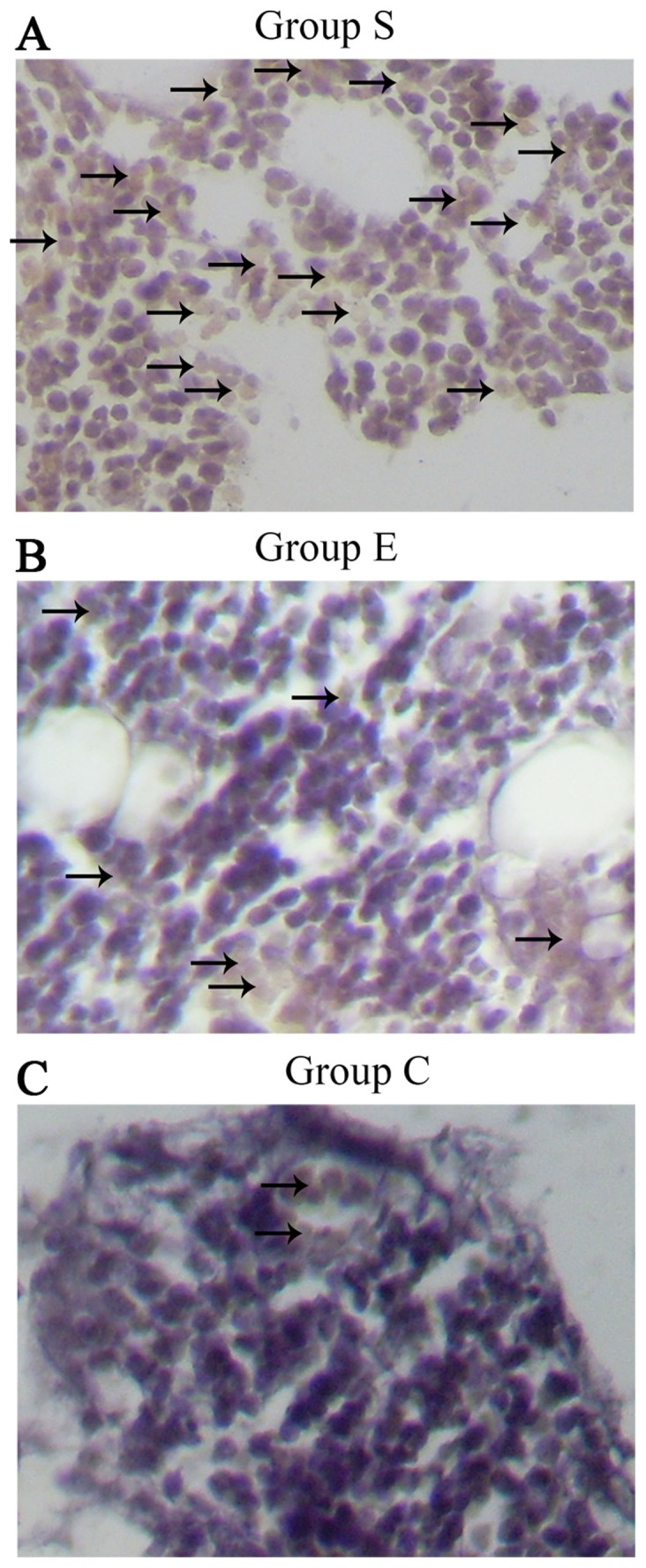
Apoptosis and DNA oxidative damage serve significant roles in the pathogenesis of steroid‑induced femoral head necrosis. Vitamin E demonstrates anti‑apoptotic and anti‑oxidant properties. Therefore, the present study investigated the effects of vitamin E on osteocyte apoptosis and DNA oxidative damage in bone marrow hemopoietic cells at an early stage of steroid‑induced femoral head osteonecrosis. Japanese white rabbits were randomly divided into three groups (steroid, vitamin E‑treated, and control groups), each comprising 12 rabbits. Those in the steroid group (group S) were initially injected twice with an intravenous dose of 100 µg/kg Escherichia coli endotoxin, with a 24 h interval between the two injections, and then with an intramuscular dose of 20 mg/kg methylprednisolone, three times at intervals of 24 h in order to establish a rabbit model of osteonecrosis. The vitamin E treated group (group E) received the same treatment as group S, and were administered 0.6 g/kg/d vitamin E daily from the beginning of modeling. The control group (group C) was injected with normal saline at the equivalent dosage and times as the aforementioned two groups. Two time points, weeks 4 and 6 following the completion of modeling, were selected. Osteonecrosis was verified by histopathology with hematoxylin-eosin staining. The apoptosis rate of osteonecrosis was analyzed by terminal deoxynucleotidyl transferase dUTP nick end labeling assay. The apoptosis expression levels of caspase‑3 and B‑cell lymphoma 2 (Bcl‑2), and DNA oxidative damage of bone marrow hematopoietic cells were analyzed by immunohistochemistry. At weeks 4 and 6 following the completion of modeling, the vacant bone lacunae rates of group E were 15.87±1.97 and 25.09±2.67%, respectively, lower than the results of 20.02±2.21 and 27.79±1.39% for group S; and the osteocyte apoptosis indexes of group E were 20.99±2.95 and 33.93±1.62%, respectively, lower than the results of 26.46±3.37 and 39.90±3.74% from group S. In addition, the Bcl-2 expression at week 4 in the femoral head tissues of group E was higher compared with group S; and the proportion of Bcl‑2‑positive cells of group E was 9.81±1.01%, higher compared with group S at 8.26±1.13%. The caspase‑3 staining data at week 4 in femoral head tissues demonstrated that in the 12 femoral heads of group S, four were negative (32%) and eight were positive (68%); in group E, five were negative (45%) and seven were positive (55%); and in group C, 11 were negative (95%) and one was positive (5%). In addition, the DNA oxidative damage rate at week 4 in the bone marrow hemopoietic cells of group E was (7.24±1.44%), lower compared with group S (11.80±1.26%), and higher compared with group C (5.75±1.47%). Vitamin E is effective in intervening in apoptosis through decreasing caspase‑3 expression and upregulating Bcl‑2 expression, and by alleviating DNA oxidative damage in bone marrow hemopoietic cells at the early stage of steroid‑induced femoral head necrosis in rabbit models.
细胞凋亡和DNA氧化损伤在类固醇诱导的股骨头坏死发病机制中起重要作用。维生素E具有抗细胞凋亡和抗氧化特性。因此,本研究调查了维生素E对类固醇诱导的股骨头坏死早期骨髓造血细胞中骨细胞凋亡和DNA氧化损伤的影响。将日本白兔随机分为三组(类固醇组、维生素E治疗组和对照组),每组12只。类固醇组(S组)最初静脉注射两次剂量为100μg/kg的大肠杆菌内毒素,两次注射间隔24小时,然后肌肉注射剂量为20mg/kg的甲基强的松龙,每隔24小时注射三次,以建立骨坏死兔模型。维生素E治疗组(E组)接受与S组相同的治疗,并从建模开始每天给予0.6g/kg/d的维生素E。对照组(C组)以与上述两组相同的剂量和时间注射生理盐水。选择建模完成后的第4周和第6周这两个时间点。通过苏木精-伊红染色的组织病理学检查验证骨坏死。通过末端脱氧核苷酸转移酶dUTP缺口末端标记法分析骨坏死的凋亡率。通过免疫组织化学分析骨髓造血细胞中半胱天冬酶-3和B细胞淋巴瘤2(Bcl-2)的凋亡表达水平以及DNA氧化损伤情况。建模完成后的第4周和第6周,E组的空骨陷窝率分别为15.87±1.97%和25.09±2.67%,低于S组的20.02±2.21%和27.79±1.39%;E组的骨细胞凋亡指数分别为20.99±2.95%和33.93±1.62%,低于S组的26.46±3.37%和39.90±3.74%。此外,E组股骨头组织在第4周时的Bcl-2表达高于S组;E组Bcl-2阳性细胞比例为9.81±1.01%,高于S组的8.26±1.13%。股骨头组织在第4周时的半胱天冬酶-3染色数据显示,S组的12个股骨头中,4个为阴性(32%),8个为阳性(68%);E组中,5个为阴性(45%),7个为阳性(55%);C组中,11个为阴性(95%),1个为阳性(5%)。此外,E组骨髓造血细胞在第4周时的DNA氧化损伤率为(7.24±1.44%),低于S组(11.80±1.26%),高于C组(5.75±1.47%)。在兔模型中,维生素E通过降低半胱天冬酶-3表达和上调Bcl-2表达有效干预细胞凋亡,并减轻类固醇诱导的股骨头坏死早期骨髓造血细胞中的DNA氧化损伤。