Keating Jane J, Runge Jeffrey J, Singhal Sunil, Nims Sarah, Venegas Ollin, Durham Amy C, Swain Gary, Nie Shuming, Low Philip S, Holt David E
Department of Surgery, Perelman School of Medicine, University of Pennsylvania, Philadelphia, Pennsylvania.
Center for Precision Surgery, Abramson Cancer Center, Perelman School of Medicine, University of Pennsylvania, Philadelphia, Pennsylvania.
Cancer. 2017 May 15;123(6):1051-1060. doi: 10.1002/cncr.30419. Epub 2016 Nov 7.
Complete tumor resection is the most important predictor of patient survival with non-small cell lung cancer. Methods for intraoperative margin assessment after lung cancer excision are lacking. This study evaluated near-infrared (NIR) intraoperative imaging with a folate-targeted molecular contrast agent (OTL0038) for the localization of primary lung adenocarcinomas, lymph node sampling, and margin assessment.
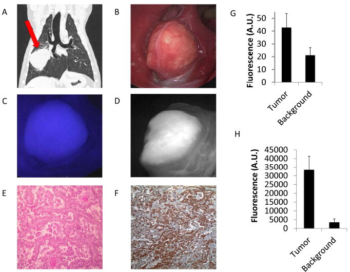
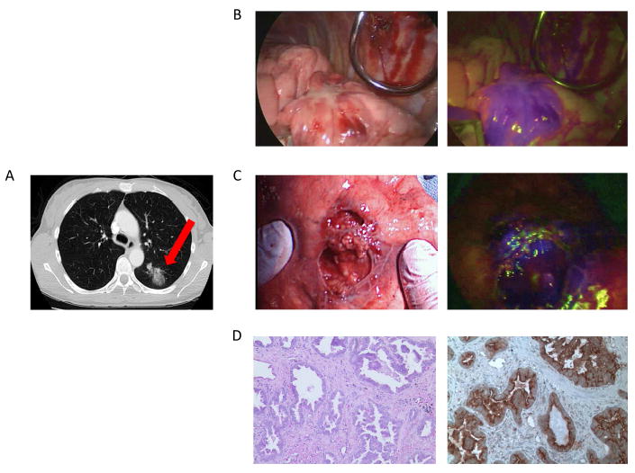
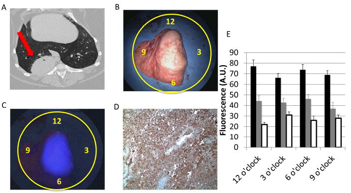
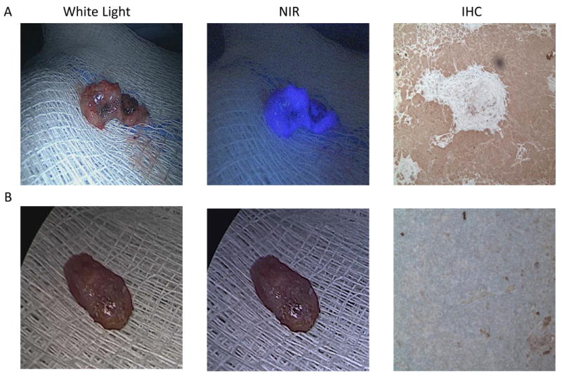
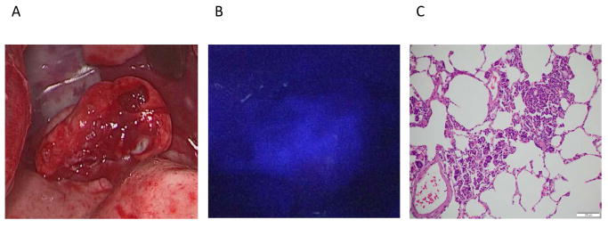
Ten dogs with lung cancer underwent either video-assisted thoracoscopic surgery or open thoracotomy and tumor excision after an intravenous injection of OTL0038. Lungs were imaged with an NIR imaging device both in vivo and ex vivo. The wound bed was re-imaged for retained fluorescence suspicious for positive tumor margins. The tumor signal-to-background ratio (SBR) was measured in all cases. Next, 3 human patients were enrolled in a proof-of-principle study. Tumor fluorescence was measured both in situ and ex vivo.
All canine tumors fluoresced in situ (mean Fluoptics SBR, 5.2 [range, 2.7-8.1]; mean Karl Storz SBR 1.9 [range, 1.4-2.6]). In addition, the fluorescence was consistent with tumor margins on pathology. Three positive lymph nodes were discovered with NIR imaging. Also, a positive retained tumor margin was discovered upon NIR imaging of the wound bed. Human pulmonary adenocarcinomas were also fluorescent both in situ and ex vivo (mean SBR, > 2.0).
NIR imaging can identify lung cancer in a large-animal model. In addition, NIR imaging can discriminate lymph nodes harboring cancer cells and also bring attention to a positive tumor margin. In humans, pulmonary adenocarcinomas fluoresce after the injection of the targeted contrast agent. Cancer 2017;123:1051-60. © 2016 American Cancer Society.
完整切除肿瘤是影响非小细胞肺癌患者生存的最重要预测因素。目前缺乏肺癌切除术中评估切缘的方法。本研究评估了使用叶酸靶向分子造影剂(OTL0038)的近红外(NIR)术中成像技术,用于原发性肺腺癌的定位、淋巴结取样及切缘评估。
10只患有肺癌的犬在静脉注射OTL0038后,接受了电视辅助胸腔镜手术或开胸手术及肿瘤切除。使用近红外成像设备对肺进行体内和体外成像。对伤口床进行再次成像,以检测残留荧光,怀疑存在阳性肿瘤切缘。在所有病例中测量肿瘤信号与背景比值(SBR)。接下来,3名人类患者参与了一项原理验证研究。对肿瘤进行原位和体外荧光测量。
所有犬类肿瘤在原位均有荧光(平均荧光光学SBR为5.2[范围2.7 - 8.1];平均卡尔·史托斯SBR为1.9[范围1.4 - 2.6])。此外,荧光与病理检查中的肿瘤切缘一致。通过近红外成像发现了3个阳性淋巴结。同时,在对伤口床进行近红外成像时发现了阳性残留肿瘤切缘。人类肺腺癌在原位和体外也有荧光(平均SBR,>2.0)。
近红外成像可在大型动物模型中识别肺癌。此外,近红外成像可鉴别含有癌细胞的淋巴结,并能发现阳性肿瘤切缘。在人类中,注射靶向造影剂后肺腺癌会发出荧光。《癌症》2017年;123:1051 - 60。©2016美国癌症协会。