Lawrimore Colleen J, Crews Fulton T
Bowles Center for Alcohol Studies , School of Medicine, University of North Carolina at Chapel Hill, Chapel Hill, North Carolina.
Curriculum in Neurobiology , University of North Carolina at Chapel Hill, Chapel Hill, North Carolina.
Alcohol Clin Exp Res. 2017 May;41(5):939-954. doi: 10.1111/acer.13368. Epub 2017 Mar 30.
Ethanol (EtOH) consumption leads to an increase of proinflammatory signaling via activation of Toll-like receptors (TLRs) such as TLR3 and TLR4 that leads to kinase activation (ERK1/2, p38, TBK1), transcription factor activation (NFκB, IRF3), and increased transcription of proinflammatory cytokines such as TNF-α, IL-1β, and IL-6. This immune signaling cascade is thought to play a role in neurodegeneration and alcohol use disorders. While microglia are considered to be the primary macrophage in brain, it is unclear what if any role neurons play in EtOH-induced proinflammatory signaling.
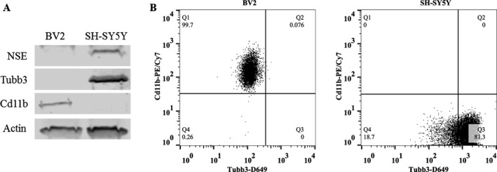
Microglia-like BV2 and retinoic acid-differentiated neuron-like SH-SY5Y were treated with TLR3 agonist Poly(I:C), TLR4 agonist lipopolysaccharide (LPS), or EtOH for 10 or 30 minutes to examine proinflammatory immune signaling kinase and transcription factor activation using Western blot, and for 24 hours to examine induction of proinflammatory gene mRNA using RT-PCR.
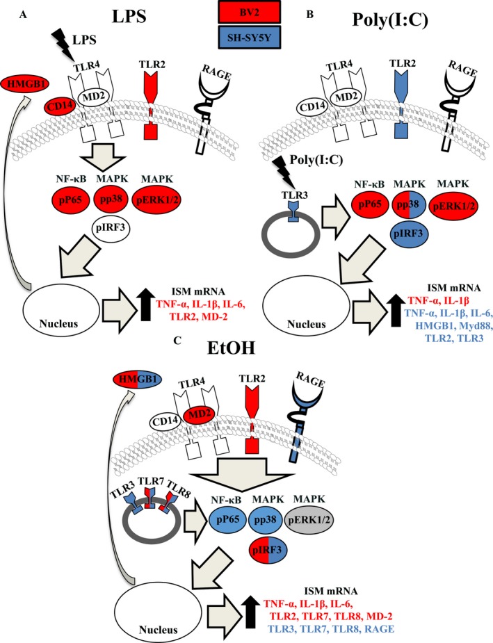
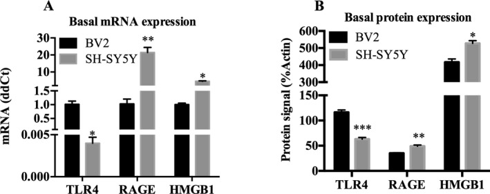
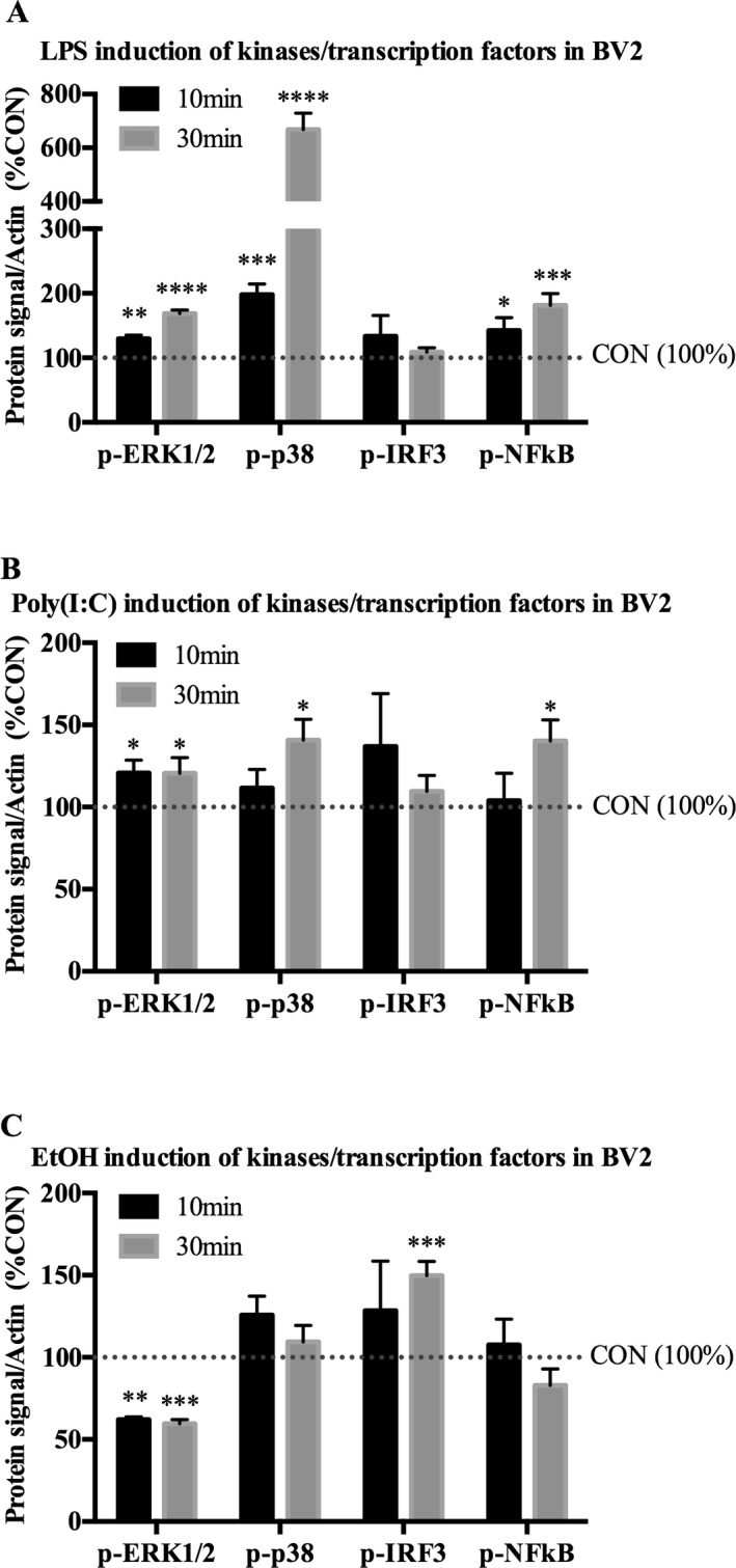
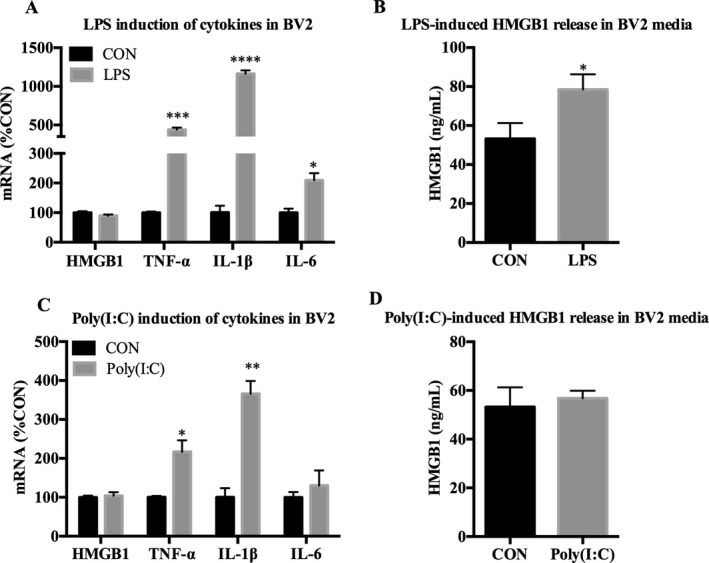
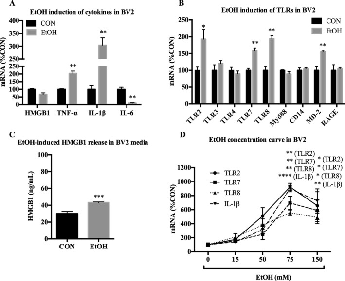
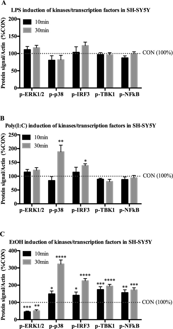
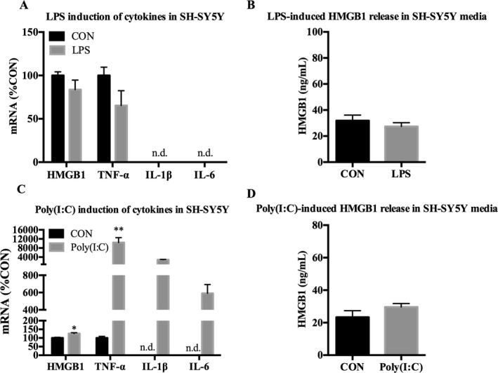
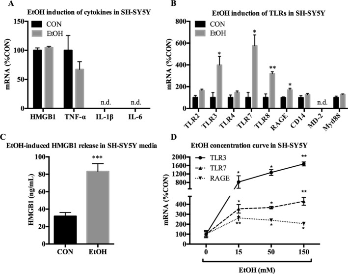
In BV2, both LPS and Poly(I:C) increased p-ERK1/2, p-p38, and p-NFκB by 30 minutes, whereas EtOH decreased p-ERK1/2 and increased p-IRF3. LPS, Poly(I:C), and EtOH all increased TNF-α and IL-1β mRNA, and EtOH further increased TLR2, 7, 8, and MD-2 mRNA in BV2. In SH-SY5Y, LPS had no effect on kinase or proinflammatory gene expression. However, Poly(I:C) increased p-p38 and p-IRF3, and increased expression of TNF-α, IL-1β, and IL-6, while EtOH increased p-p38, p-IRF3, p-TBK1, and p-NFκB while decreasing p-ERK1/2 and increasing expression of TLR3, 7, 8, and RAGE mRNA. HMGB1, a TLR agonist, was induced by LPS in BV2 and by EtOH in both cell types. EtOH was more potent at inducing proinflammatory gene mRNA in SH-SY5Y compared with BV2.
These results support a novel and unique mechanism of EtOH, TLR3, and TLR4 signaling in neuron-like SH-SY5Y and microglia-like BV2 that likely contributes to the complexity of brain neuroimmune signaling.
乙醇(EtOH)的摄入通过激活Toll样受体(TLR)如TLR3和TLR4导致促炎信号增加,进而导致激酶激活(ERK1/2、p38、TBK1)、转录因子激活(NFκB、IRF3)以及促炎细胞因子如TNF-α、IL-1β和IL-6的转录增加。这种免疫信号级联反应被认为在神经退行性变和酒精使用障碍中起作用。虽然小胶质细胞被认为是大脑中的主要巨噬细胞,但尚不清楚神经元在乙醇诱导的促炎信号中是否发挥作用以及发挥何种作用。
用TLR3激动剂聚肌胞苷酸(Poly(I:C))、TLR4激动剂脂多糖(LPS)或乙醇处理小胶质细胞样BV2和视黄酸分化的神经元样SH-SY5Y细胞10或30分钟,通过蛋白质免疫印迹法检测促炎免疫信号激酶和转录因子的激活情况,并处理24小时,通过逆转录聚合酶链反应(RT-PCR)检测促炎基因mRNA的诱导情况。
在BV2细胞中,LPS和Poly(I:C)在30分钟时均增加了p-ERK1/2、p-p38和p-NFκB的表达,而乙醇降低了p-ERK1/2的表达并增加了p-IRF3的表达。LPS、Poly(I:C)和乙醇均增加了TNF-α和IL-1β mRNA的表达,并且乙醇进一步增加了BV2细胞中TLR2、7、8和MD-2 mRNA的表达。在SH-SY5Y细胞中,LPS对激酶或促炎基因表达没有影响。然而,Poly(I:C)增加了p-p38和p-IRF3的表达,并增加了TNF-α、IL-1β和IL-6的表达,而乙醇增加了p-p38、p-IRF3、p-TBK1和p-NFκB的表达,同时降低了p-ERK1/2的表达,并增加了TLR3、7、8和RAGE mRNA的表达。高迁移率族蛋白B1(HMGB1)作为一种TLR激动剂,在BV2细胞中可被LPS诱导,在两种细胞类型中均可被乙醇诱导。与BV2细胞相比,乙醇在诱导SH-SY5Y细胞促炎基因mRNA表达方面更有效。
这些结果支持了乙醇、TLR3和TLR4信号在神经元样SH-SY5Y细胞和小胶质细胞样BV2细胞中的一种新的独特机制,这可能导致大脑神经免疫信号的复杂性。