Zhu Yu, Wang Yuchen, Zhao Bizeng, Niu Xin, Hu Bin, Li Qing, Zhang Juntao, Ding Jian, Chen Yunfeng, Wang Yang
Department of Orthopedic Surgery, Shanghai Jiao Tong University Affiliated Sixth People's Hospital, 600 Yishan Road, Shanghai, 200233, China.
Institute of Microsurgery on Extremities, Shanghai Jiao Tong University Affiliated Sixth People's Hospital, 600 Yishan Road, Shanghai, 200233, China.
Stem Cell Res Ther. 2017 Mar 9;8(1):64. doi: 10.1186/s13287-017-0510-9.
Osteoarthritis (OA) is the most common joint disease worldwide. In the past decade, mesenchymal stem cells (MSCs) have been used widely for the treatment of OA. A potential mechanism of MSC-based therapies has been attributed to the paracrine secretion of trophic factors, in which exosomes may play a major role. In this study, we aimed to compare the effectiveness of exosomes secreted by synovial membrane MSCs (SMMSC-Exos) and exosomes secreted by induced pluripotent stem cell-derived MSCs (iMSC-Exos) on the treatment of OA.
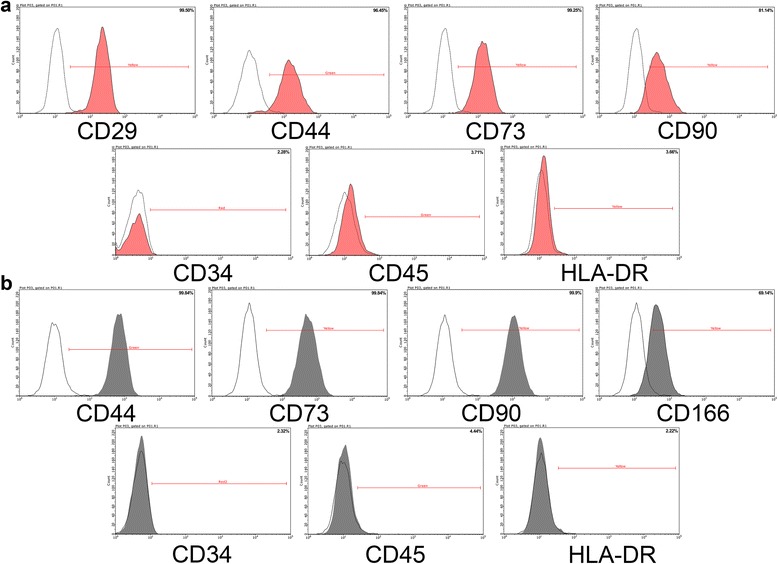
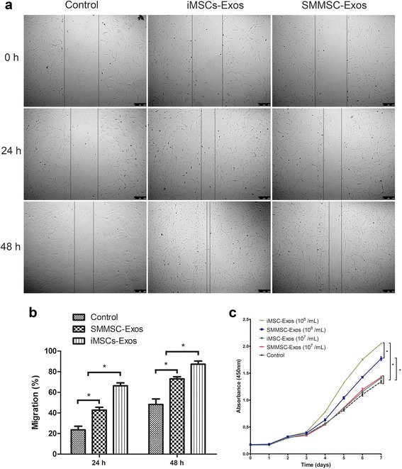
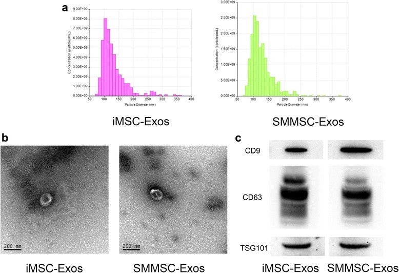
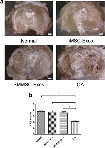
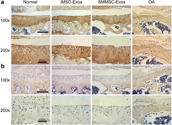
Induced pluripotent stem cell-derived MSCs and synovial membrane MSCs were characterized by flow cytometry. iMSC-Exos and SMMSC-Exos were isolated using an ultrafiltration method. Tunable resistive pulse-sensing analysis, transmission electron microscopy, and western blots were used to identify exosomes. iMSC-Exos and SMMSC-Exos were injected intra-articularly in a mouse model of collagenase-induced OA and the efficacy of exosome injections was assessed by macroscopic, histological, and immunohistochemistry analysis. We also evaluated the effects of iMSC-Exos and SMMSC-Exos on proliferation and migration of human chondrocytes by cell-counting and scratch assays, respectively.
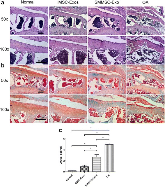
The majority of iMSC-Exos and SMMSC-Exos were approximately 50-150 nm in diameter and expressed CD9, CD63, and TSG101. The injection of iMSC-Exos and SMMSC-Exos both attenuated OA in the mouse OA model, but iMSC-Exos had a superior therapeutic effect compared with SMMSC-Exos. Similarly, chondrocyte migration and proliferation were stimulated by both iMSC-Exos and SMMSC-Exos, with iMSC-Exos exerting a stronger effect.
The present study demonstrated that iMSC-Exos have a greater therapeutic effect on OA than SMMSC-Exos. Because autologous iMSCs are theoretically inexhaustible, iMSC-Exos may represent a novel therapeutic approach for the treatment of OA.
骨关节炎(OA)是全球最常见的关节疾病。在过去十年中,间充质干细胞(MSCs)已被广泛用于OA的治疗。基于MSCs治疗的一种潜在机制归因于营养因子的旁分泌,其中外泌体可能起主要作用。在本研究中,我们旨在比较滑膜间充质干细胞分泌的外泌体(SMMSC-Exos)和诱导多能干细胞来源的间充质干细胞分泌的外泌体(iMSC-Exos)对OA治疗的有效性。
通过流式细胞术对诱导多能干细胞来源的间充质干细胞和滑膜间充质干细胞进行表征。使用超滤方法分离iMSC-Exos和SMMSC-Exos。采用可调电阻脉冲传感分析、透射电子显微镜和蛋白质免疫印迹法鉴定外泌体。将iMSC-Exos和SMMSC-Exos关节内注射到胶原酶诱导的OA小鼠模型中,并通过宏观、组织学和免疫组织化学分析评估外泌体注射的疗效。我们还分别通过细胞计数和划痕试验评估了iMSC-Exos和SMMSC-Exos对人软骨细胞增殖和迁移的影响。
大多数iMSC-Exos和SMMSC-Exos直径约为50-150nm,并表达CD9、CD63和TSG101。在小鼠OA模型中,注射iMSC-Exos和SMMSC-Exos均减轻了OA,但与SMMSC-Exos相比,iMSC-Exos具有更好的治疗效果。同样,iMSC-Exos和SMMSC-Exos均刺激了软骨细胞的迁移和增殖,且iMSC-Exos的作用更强。
本研究表明,iMSC-Exos对OA的治疗效果优于SMMSC-Exos。由于自体iMSCs理论上是取之不尽的,iMSC-Exos可能代表一种治疗OA的新型治疗方法。