Fuentes-Santamaría Verónica, Alvarado Juan Carlos, Melgar-Rojas Pedro, Gabaldón-Ull María C, Miller Josef M, Juiz José M
Instituto de Investigación en Discapacidades NeurológicasAlbacete, Spain; Facultad de Medicina, Universidad de Castilla-La ManchaAlbacete, Spain.
Center for Hearing and Communication Research and Department of Clinical Neuroscience, Karolinska InstitutetStockholm, Sweden; Kresge Hearing Research Institute, University of MichiganAnn Arbor, MI, USA.
Front Neuroanat. 2017 Feb 23;11:9. doi: 10.3389/fnana.2017.00009. eCollection 2017.
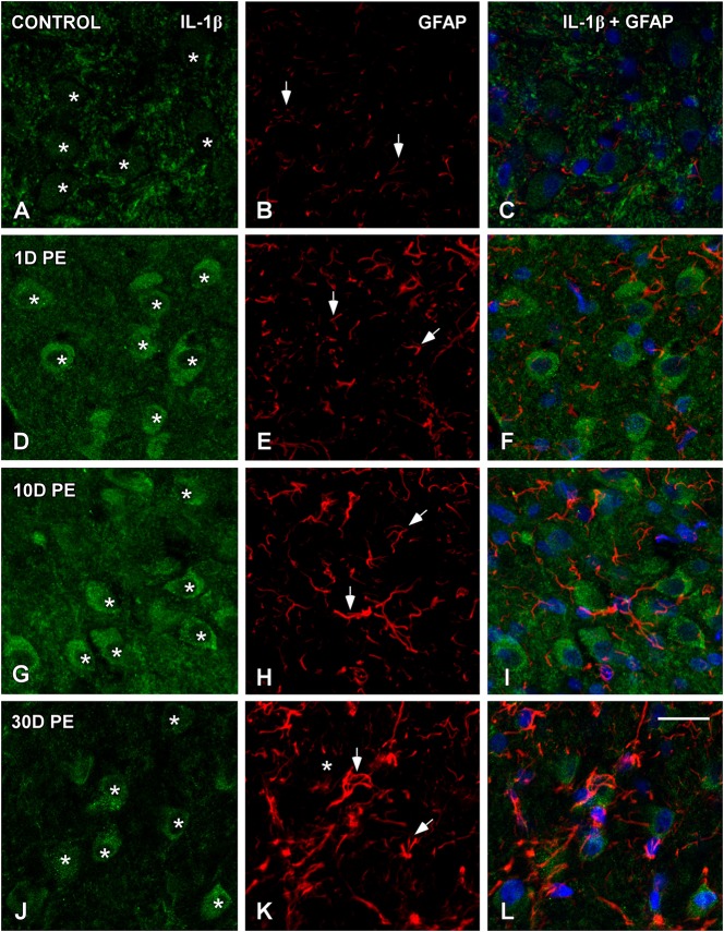
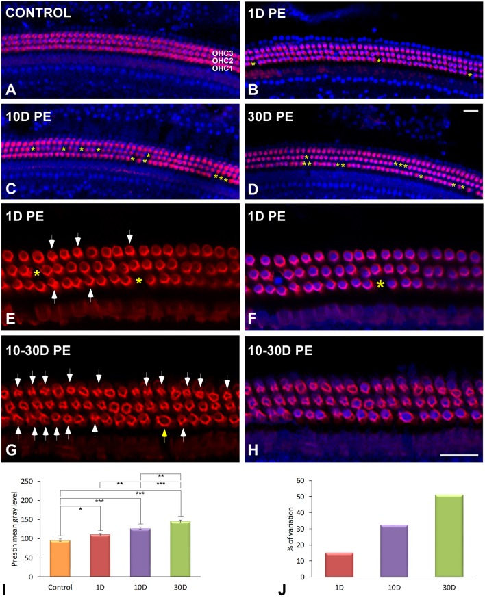
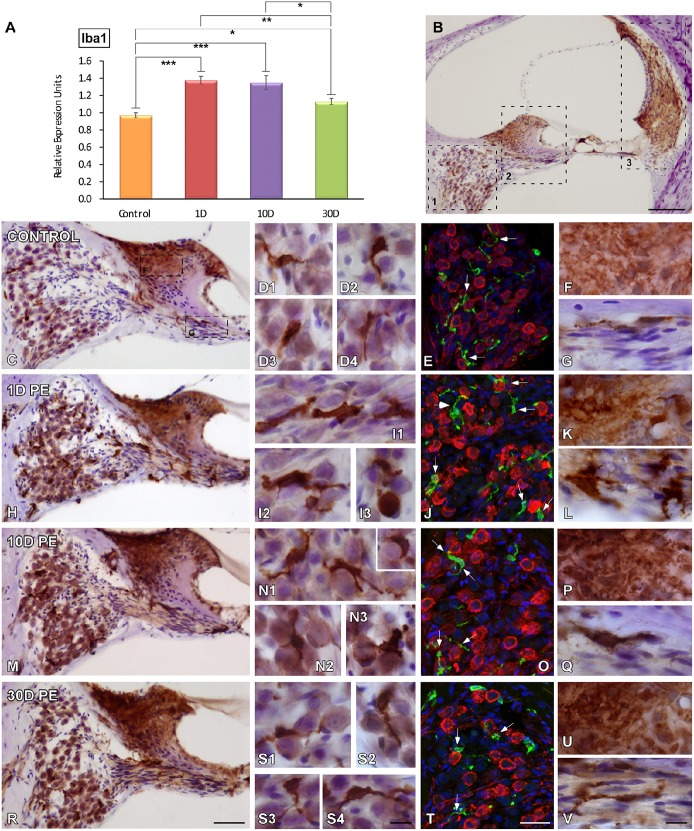
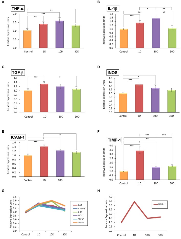
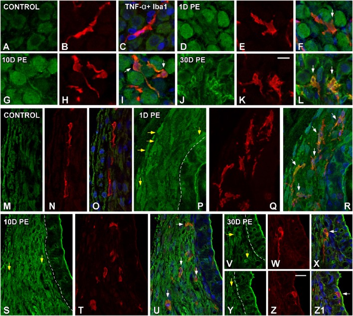
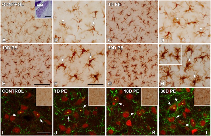
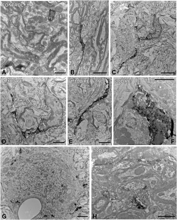
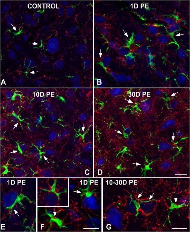
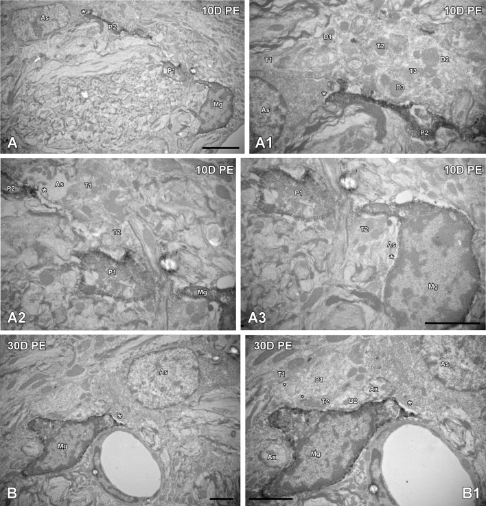
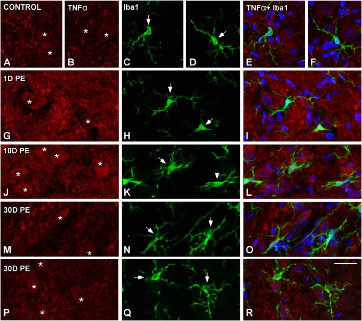
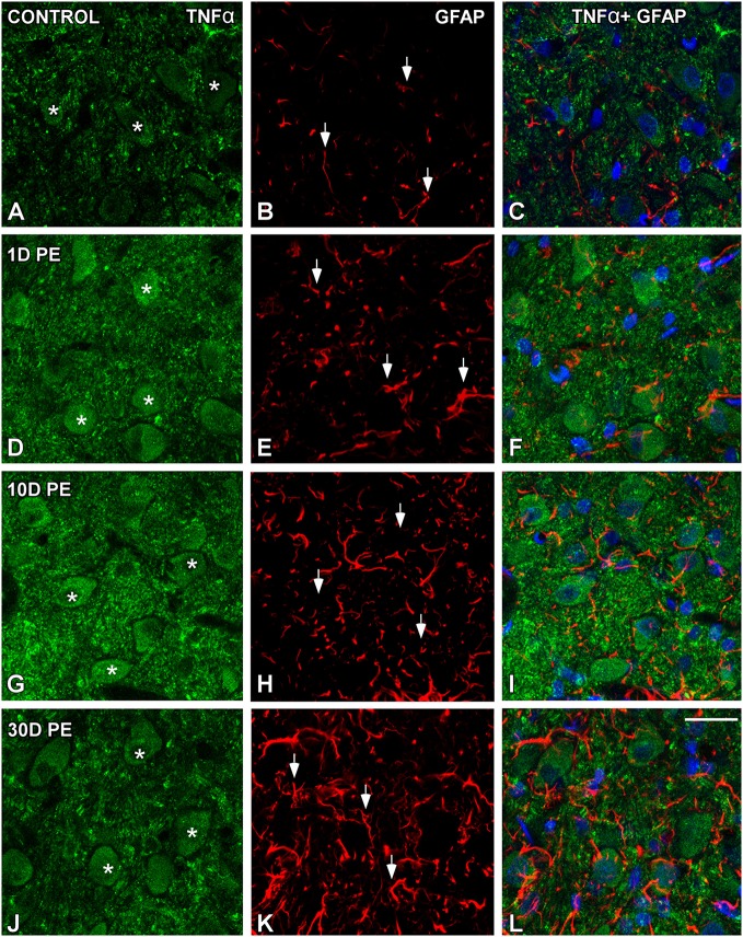
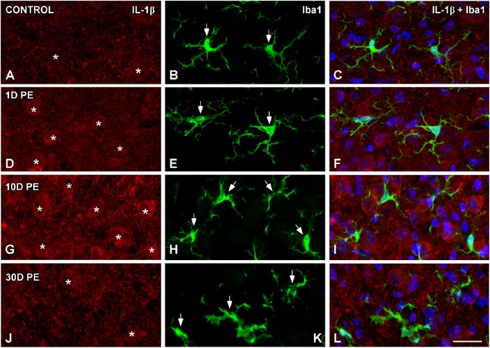
Repeated noise exposure induces inflammation and cellular adaptations in the peripheral and central auditory system resulting in pathophysiology of hearing loss. In this study, we analyzed the mechanisms by which noise-induced inflammatory-related events in the cochlea activate glial-mediated cellular responses in the cochlear nucleus (CN), the first relay station of the auditory pathway. The auditory function, glial activation, modifications in gene expression and protein levels of inflammatory mediators and ultrastructural changes in glial-neuronal interactions were assessed in rats exposed to broadband noise (0.5-32 kHz, 118 dB SPL) for 4 h/day during 4 consecutive days to induce long-lasting hearing damage. Noise-exposed rats developed a permanent threshold shift which was associated with hair cell loss and reactive glia. Noise-induced microglial activation peaked in the cochlea between 1 and 10D post-lesion; their activation in the CN was more prolonged reaching maximum levels at 30D post-exposure. RT-PCR analyses of inflammatory-related genes expression in the cochlea demonstrated significant increases in the mRNA expression levels of pro- and anti-inflammatory cytokines, inducible nitric oxide synthase, intercellular adhesion molecule and tissue inhibitor of metalloproteinase-1 at 1 and 10D post-exposure. In noise-exposed cochleae, interleukin-1β (IL-1β), and tumor necrosis factor α (TNF-α) were upregulated by reactive microglia, fibrocytes, and neurons at all time points examined. In the CN, however, neurons were the sole source of these cytokines. These observations suggest that noise exposure causes peripheral and central inflammatory reactions in which TNF-α and IL-1β are implicated in regulating the initiation and progression of noise-induced hearing loss.
反复暴露于噪声会诱发外周和中枢听觉系统的炎症及细胞适应性变化,从而导致听力损失的病理生理学改变。在本研究中,我们分析了耳蜗中噪声诱导的炎症相关事件激活听觉通路第一中继站耳蜗核(CN)中胶质细胞介导的细胞反应的机制。对连续4天每天暴露于宽带噪声(0.5 - 32 kHz,118 dB SPL)4小时以诱导长期听力损伤的大鼠,评估其听觉功能、胶质细胞激活、炎症介质基因表达和蛋白质水平的改变以及胶质细胞与神经元相互作用的超微结构变化。噪声暴露大鼠出现了永久性阈移,这与毛细胞损失和反应性胶质细胞有关。噪声诱导的小胶质细胞激活在损伤后1至10天在耳蜗中达到峰值;它们在CN中的激活持续时间更长,在暴露后30天达到最高水平。对耳蜗中炎症相关基因表达的RT-PCR分析表明,暴露后1天和10天,促炎和抗炎细胞因子、诱导型一氧化氮合酶、细胞间粘附分子和金属蛋白酶组织抑制剂-1的mRNA表达水平显著增加。在噪声暴露的耳蜗中,在所有检测时间点,反应性小胶质细胞、纤维细胞和神经元均上调白细胞介素-1β(IL-1β)和肿瘤坏死因子α(TNF-α)。然而,在CN中,神经元是这些细胞因子的唯一来源。这些观察结果表明,噪声暴露会引起外周和中枢炎症反应,其中TNF-α和IL-1β参与调节噪声诱导的听力损失的起始和进展。