Chen Yu-Chen, Chen Guang-Di, Auerbach Benjamin D, Manohar Senthilvelan, Radziwon Kelly, Salvi Richard
Department of Radiology, Nanjing First Hospital, Nanjing Medical University, 210006 Nanjing, China; Center for Hearing and Deafness, SUNY at Buffalo, Buffalo, NY 14214, USA.
Center for Hearing and Deafness, SUNY at Buffalo, Buffalo, NY 14214, USA.
Hear Res. 2017 Jun;349:208-222. doi: 10.1016/j.heares.2017.03.005. Epub 2017 Mar 7.
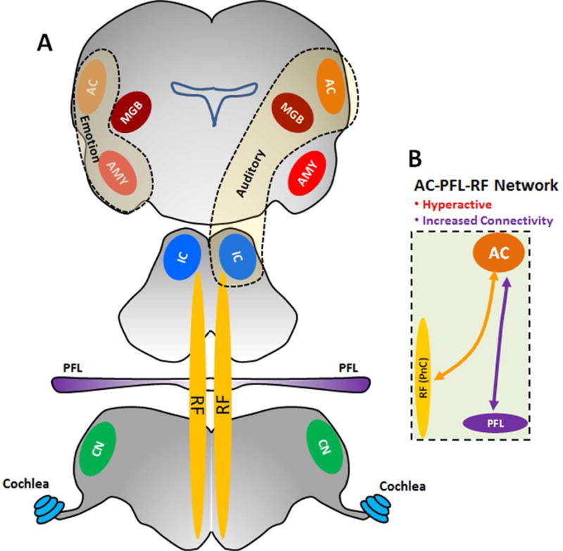
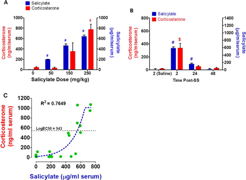
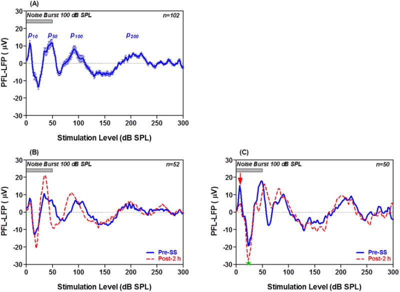
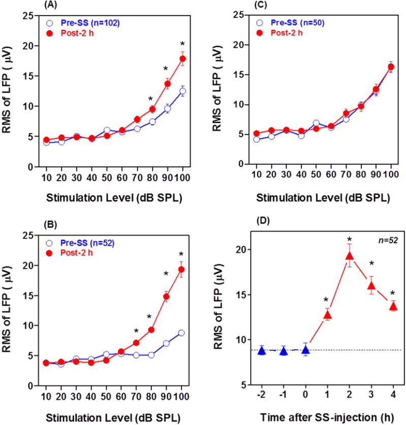
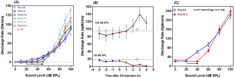
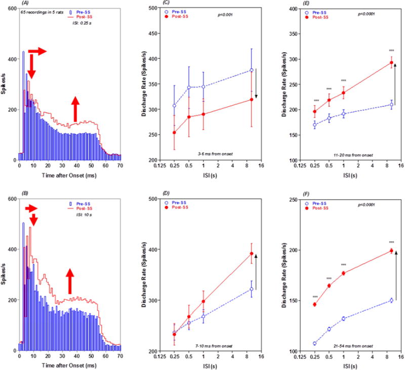
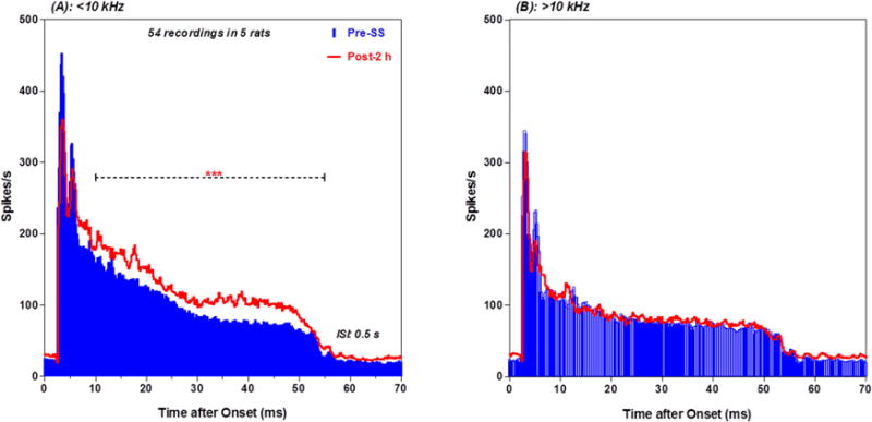
Tinnitus and hyperacusis are common and potentially serious hearing disorders associated with noise-, age- or drug-induced hearing loss. Accumulating evidence suggests that tinnitus and hyperacusis are linked to excessive neural activity in a distributed brain network that not only includes the central auditory pathway, but also brain regions involved in arousal, emotion, stress and motor control. Here we examine electrophysiological changes in two novel non-auditory areas implicated in tinnitus and hyperacusis: the caudal pontine reticular nucleus (PnC), involved in arousal, and the paraflocculus lobe of the cerebellum (PFL), implicated in head-eye coordination and gating tinnitus and we measure the changes in corticosterone stress hormone levels. Using the salicylate-induced model of tinnitus and hyperacusis, we found that long-latency (>10 ms) sound-evoked response components in both the brain regions were significantly enhanced after salicylate administration, while the short-latency responses were reduced, likely reflecting cochlear hearing loss. These results are consistent with the central gain model of tinnitus and hyperacusis, which proposes that these disorders arise from the amplification of neural activity in central auditory pathway plus other regions linked to arousal, emotion, tinnitus gating and motor control. Finally, we demonstrate that salicylate results in an increase in corticosterone level in a dose-dependent manner consistent with the notion that stress may interact with hearing loss in tinnitus and hyperacusis development. This increased stress response has the potential to have wide-ranging effects on the central nervous system and may therefore contribute to brain-wide changes in neural activity.
耳鸣和听觉过敏是常见且可能严重的听力障碍,与噪声、年龄或药物引起的听力损失有关。越来越多的证据表明,耳鸣和听觉过敏与分布式脑网络中的过度神经活动有关,该网络不仅包括中枢听觉通路,还包括参与觉醒、情绪、压力和运动控制的脑区。在这里,我们研究了两个与耳鸣和听觉过敏有关的新的非听觉区域的电生理变化:参与觉醒的尾侧脑桥网状核(PnC)和参与头眼协调以及耳鸣门控的小脑旁绒球叶(PFL),并测量了皮质酮应激激素水平的变化。使用水杨酸盐诱导的耳鸣和听觉过敏模型,我们发现,给药后,这两个脑区中长潜伏期(>10毫秒)的声音诱发反应成分均显著增强,而短潜伏期反应则减弱,这可能反映了耳蜗性听力损失。这些结果与耳鸣和听觉过敏的中枢增益模型一致,该模型提出,这些障碍源于中枢听觉通路以及与觉醒、情绪、耳鸣门控和运动控制相关的其他区域的神经活动放大。最后,我们证明水杨酸盐会导致皮质酮水平以剂量依赖性方式升高,这与压力可能在耳鸣和听觉过敏发展过程中与听力损失相互作用的观点一致。这种增加的应激反应有可能对中枢神经系统产生广泛影响,因此可能导致全脑神经活动的变化。