Ordeix Laura, Dalmau Annabel, Osso Montsant, Llull Joan, Montserrat-Sangrà Sara, Solano-Gallego Laia
Departament de Medicina i Cirurgia Animals, Facultat de Veterinària, Universitat Autònoma de Barcelona, Bellaterra, Spain.
Hospital Clínic Veterinari, Universitat Autònoma de Barcelona, Bellaterra, Spain.
Parasit Vectors. 2017 Mar 13;10(1):121. doi: 10.1186/s13071-017-2051-6.
Normal-looking skin of dogs with leishmaniosis frequently shows microscopic lesions along with the presence of Leishmania amastigotes. However, histological lesions with or without detection of amastigotes might not occur in less severe clinical cases. In addition, comparative studies between paired clinically-lesioned and normal-looking skin samples from dogs with different disease severity are lacking. The objective of this study was to compare histological and parasitological findings by Leishmania immunohistochemistry (IHC) and quantitative PCR (qPCR) on paired clinically-lesioned and normal-looking skin biopsies from 25 dogs with different clinical stages of leishmaniosis, 11 with stage I-mild disease (papular dermatitis) and 14 with stage II-III (ulcerative or exfoliative dermatitis).
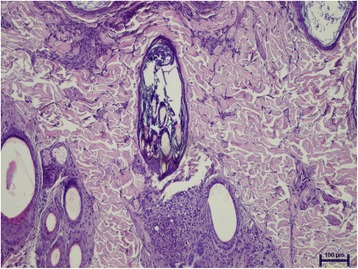
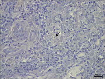
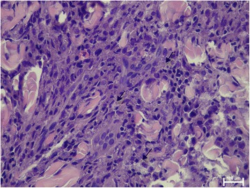
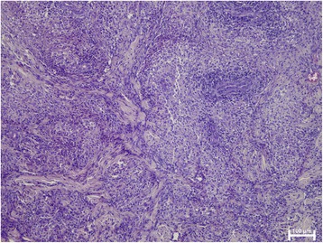
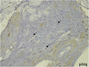
The study demonstrated microscopic lesions in 14 out of 25 (56%) samples from normal-looking skin biopsies. In those samples, perivascular to interstitial dermatitis composed by macrophages with lymphocytes and plasma cells was observed mainly in the superficial and mid-dermis. The intensity of the dermatitis was mild to moderate and always less prominent than in the clinically-lesioned skin. In normal-looking skin samples, the presence of parasites was detected by histology, IHC and qPCR in 5/25 (20%), 8/25 (32%) and 18/25 (72%), respectively. Leishmania was encountered in 11/25 (44%), 23/25 (92%) and 25/25 (100%) of clinically-lesioned skin samples by histology, IHC and qPCR, respectively. Normal-looking skin from dogs with stage I-mild disease was less frequently inflamed (P = 0.0172). Furthermore, Leishmania was more easily demonstrated by histology (P = 0.0464), IHC (P = 0.0421) or qPCR (P = 0.0068) in normal-looking skin of dogs with stage II-III-moderate to severe disease. In addition, in the latter group, there was a significantly higher parasite load studied by means of qPCR than in dogs with less severe disease (P = 0.043). Clinically-lesioned skin from dogs with stage I disease was more frequently characterised by the nodular to diffuse pattern and granuloma formation (P = 0.0166) and by a lower parasite load studied by means of qPCR (P = 0.043) compared with more diseased dogs.
Normal-looking skin from dogs with stage I is less likely to present histological lesions as well as harbour the parasite when compared with dogs with moderate to severe leishmaniosis.
患有利什曼病的犬只外观正常的皮肤通常会出现微观病变,并伴有利什曼原虫无鞭毛体。然而,在病情较轻的临床病例中,可能不会出现有或没有检测到无鞭毛体的组织学病变。此外,目前缺乏对不同疾病严重程度的犬只配对的临床病变皮肤样本和外观正常皮肤样本进行的比较研究。本研究的目的是通过利什曼原虫免疫组织化学(IHC)和定量聚合酶链反应(qPCR),比较25只处于利什曼病不同临床阶段的犬只配对的临床病变皮肤活检样本和外观正常皮肤活检样本的组织学和寄生虫学结果,其中11只处于I期轻度疾病(丘疹性皮炎),14只处于II - III期(溃疡性或剥脱性皮炎)。
该研究在25份外观正常的皮肤活检样本中的14份(56%)中发现了微观病变。在这些样本中,主要在浅表和真皮中层观察到由巨噬细胞、淋巴细胞和浆细胞组成的血管周围至间质性皮炎。皮炎的强度为轻度至中度,且总是不如临床病变皮肤明显。在外观正常的皮肤样本中,通过组织学、免疫组织化学和定量聚合酶链反应分别在5/25(20%)、8/25(32%)和18/25(72%)中检测到寄生虫的存在。通过组织学、免疫组织化学和定量聚合酶链反应分别在11/25(44%)、23/25(92%)和25/25(100%)的临床病变皮肤样本中检测到利什曼原虫。I期轻度疾病犬只的外观正常皮肤炎症较少见(P = 0.0172)。此外,在II - III期中度至重度疾病犬只的外观正常皮肤中,通过组织学(P = 0.0464)、免疫组织化学(P = 0.0421)或定量聚合酶链反应(P = 0.0068)更容易检测到利什曼原虫。此外,在后者组中,通过定量聚合酶链反应研究的寄生虫载量明显高于病情较轻的犬只(P = 0.043)。与病情较重的犬只相比,I期疾病犬只的临床病变皮肤更常表现为结节状至弥漫性模式和肉芽肿形成(P = 0.0166),且通过定量聚合酶链反应研究的寄生虫载量较低(P = 0.043)。
与中度至重度利什曼病犬只相比,I期犬只外观正常的皮肤出现组织学病变以及携带寄生虫的可能性较小。